Benzinger Lab
Projects
The Benzinger Lab has a number of ongoing projects that are recruiting at any given time. Below is an overview of what we are working on at the moment. To see our currently open clinical trials, visit ClinicalTrials.gov.
New Imaging Techniques
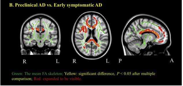
Alzheimer disease (AD) is preceded by at least a decade of clinically silent brain changes (termed “preclinical AD”) that ultimately result in declines in memory and thinking. Inflammation in the brain plays an important role neurodegenerative diseases, particularly in the transition from normal cognition to dementia, but we lack easily accessible tools to identify neuroinflammation. These projects use a novel brain MR imaging test, diffusion basis spectrum imaging (DBSI), and PET study with 11C-CS1P1 to examine inflammation during the stages of preclinical and clinical Alzheimer’s disease and also in multiple sclerosis.
Funding
1R01AG054567-01A1
2R01NS103988-06A1
Collaborators
Yong Wang, PhD (Co-Principal Investigator)
Beau Ances, MD, PhD
Matthew Brier, MD, PhD
Anne Cross, MD
Richard Perrin, MD, PhD
Suzanne Schindler, MD, PhD
Zhude Tu, PhD
Qing Wang, PhD
Jinbin Xu, PhD
This project is focused upon a bench-to-bedside translation of a molecular imaging PET radiotracer for noninvasive interrogation of Alzheimer’s disease (AD) at prodromal stages. Following validation in humans, the agent could also be useful for noninvasive interrogation of role of Aβ pathophysiology in traumatic brain injury (TBI), chronic traumatic encephalopathy (CTE) and differentiation of Parkinson’s disease patients with dementia (PDD) from their counterparts only with Parkinson’s disease (PD).
Funding
1RF1AG064937-01
The lab serves as the imaging core for the Knight Alzheimer Disease Research Center, the international studies of ADAD, the Dominantly Inherited Alzheimer Network (DIAN) and the DIAN-Therapeutic Unit. (Gordon et al., Brain, 2019)
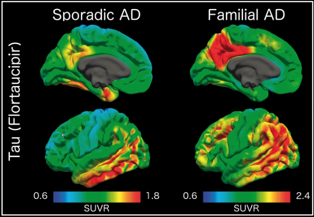
Tauopathy is a hallmark pathology of Alzheimer’s disease (AD) with a strong relationship with cognitive impairment. Tau PET uptake is tied to the onset of cognitive dysfunction, and there is a higher amount, and different regional pattern of binding in autosomal dominant AD compared to late onset, non-familial AD.
Funding
- ADRC Project 1, NIH/NIA P50AG05681
- Avid Radiopharmaceuticals (a wholly-owned subsidiary of Eli Lilly and Company) provided technology transfer and precursor for AV-1451
- DIAN scans also supported by R01AG05255003
- Driving Performance in Preclinical AD, NIH R01AG043434
- HASD Project 4, NIH/NIA P01AG003991
- Hope Center for Neurological Disorders
- Institute of Clinical and Translational Sciences (ICTS) at Washington University (posterior cortical atrophy cohort support)
- ICTS NIH/NCATS UL1TR000448 and Barnes-Jewish Hospital Foundation (DIAN WU scans support)
- LEFTS (FTD cohort support)
- McDonnell Center for Systems Neuroscience (ALS cohort support)
- NIH R01NR014449 (HIV cohort support)
- Willman Scholar Fund (BJHF)
Collaborators
Beau Ances, MD, PhD (Co-Principal Investigator)
Brian Gordon, PhD
Knight Alzheimer Research Imaging (KARI) Program
The lab serves as the imaging core for the ADRC, the international studies of ADAD, the Dominantly Inherited Alzheimer Network (DIAN) and the DIAN-Therapeutic Unit.
Funding
- ADRC Center Grant, NIH/NIA P50AG005681
- Charles and Joanne Knight Alzheimer Disease Research Center Support Fund
- The Foundation for Barnes-Jewish Hospital
- David and Betty Farrell Medical Research Fund
- Daniel J Brennan Alzheimer Research Fund
- Fred Simmons and Olga Mohan Alzheimer Research Support Fund
- Thomas E. Brew Foundation Fund
- Willman Scholar Fund (BJHF)
The ACS is a longitudinal study of aging, beginning at age 45. Imaging includes MRI, amyloid PIB-PET, and tau T807 PET. Early structural and functional changes in the brains of individuals at risk for developing dementia of the Alzheimer type (DAT) are not well understood. Typically a diagnosis of DAT is made using neuropsychological performance testing and clinical evaluation. Previous work by our group and others has identified a preclinical phase of DAT that is associated with a constellation of changes in biomarkers including decreased cerebral spinal fluid (CSF) Abeta42 (AP42) levels, increased fibrillar amyloid deposits in the brain as detected by PET.
Funding
- IH/NIA P01AG026276
- Fred Simmons and Olga Mohan (Should this be: Fred Simmons and Olga Mohan Alzheimer Research Support Fund)
- Test-retest supported by ICTS-JIT = CTS ID#: JIT172M & MIR ID#: MIR11-022J
- The McDonnell Center for Systems Neuroscience, 22 3922 26239N
Collaborators
Manu Goyal, MD, Andrei Vlassenko, MD, PhD, and Marcus Raichle, MD, are performing [15]O and FDG metabolic PET in this cohort.
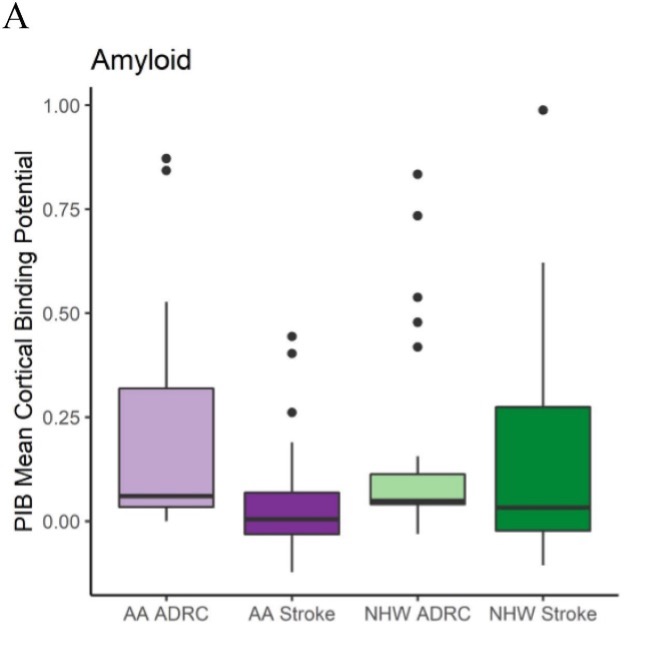
This is a longitudinal study of aging, beginning at age 65. Imaging includes MRI, amyloid AV45-PET, and tau T807 (18F-AV-1451) PET.
Stroke and Alzheimer’s disease share risk factors and often co-occur, and both have been reported to have a higher prevalence in African Americans as compared to non-Hispanic whites. In a study of 243 participants, of which 52% identified as African Americans, we found that Amyloid PET not different for African American compared to non-Hispanic whites. (Koenig et al., NeuroImage: Clinical, 2021)
Funding
- Avid Radiopharmaceuticals, a wholly owned subsidiary of Eli Lilly (florbetapir and AV1451 imaging). For Florbetapir (AV45), Avid provided the doses and partial support for scanning through an investigator-initiated research grant awarded to Washington University (J.C. Morris, T.L.S. Benzinger).
- Barnes-Jewish Hospital Foundation (PIB imaging)
- Mallinckrodt Institute of Radiology (MRI in participants with dementia)
- NIH/NIA P01AG003991
International Collaborations for Autosomal Dominant AD
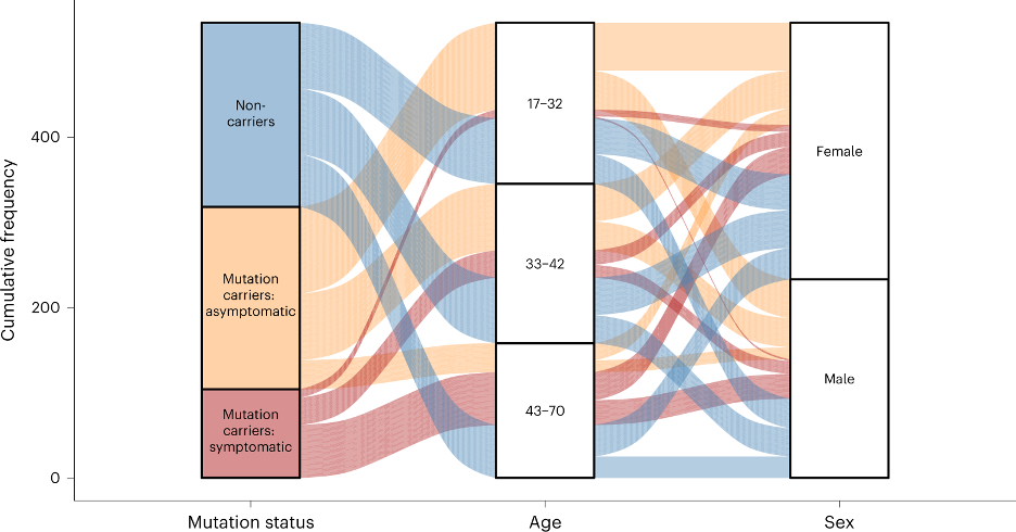
Dominantly Inherited Alzheimer Network (DIAN) Imaging Core Lab
An international study of families with autosomal dominant AD. Imaging includes MRI, amyloid PIB and AV45 PET, and tau T807 PET.
- NIA/NINDA U01AG042791-02 Phase II
- NIH/NIA UF1AG032438, NIH/NIA U19AG03243808 and the German Center for Neurodegenerative Diseases (DZNE)
- NIH/NIA R01AG052550-01A1 for Tau PET in DIAN, and imaging-pathology correlation studies
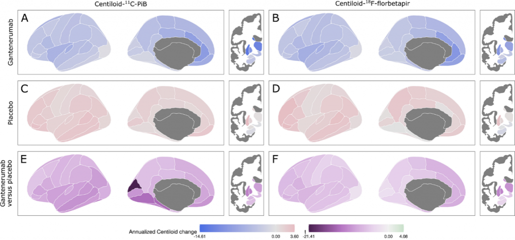
Dominantly Inherited Alzheimer Network Trials Unit (DIAN-TU) Imaging Core Lab
An international study of families with autosomal dominant AD. Imaging includes MRI, amyloid PIB and AV45 PET, and tau T807 (AV-1451) PET.
- NIA/NINDA U01AG042791-02 Phase II
- NIH/NIA R01AG046179 Phase III
- DIAN Trials Pharma Consortium
- Avid Radiopharmaceuticals, Lilly, Roche, MNIWillman Scholar Fund (BJHF)
Clinical Translational Research: Brain Tumors
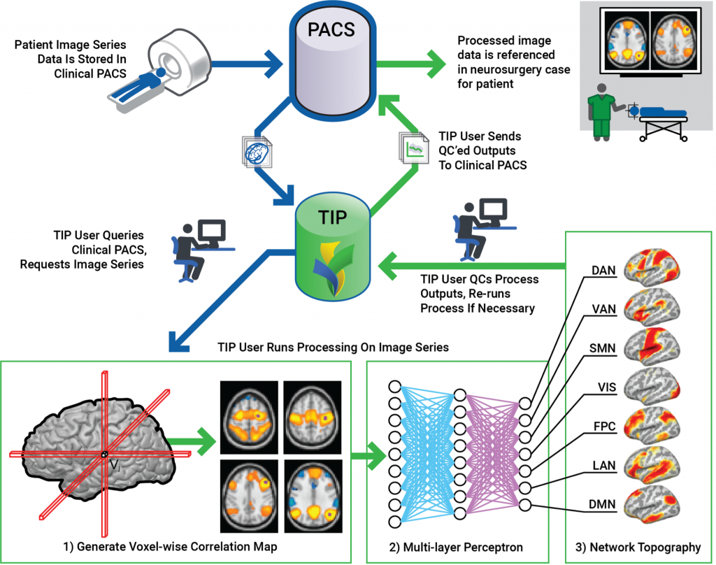
Neurosurgical Translational Interface Pipeline for Clinical Resting State Functional MRI
The goal of this project is to develop a clinical workflow for volumetric MRI and resting state functional MRI for preoperative planning and for evaluation of dementia.
Additional Sources of Funding
- Willman Scholar, The Foundation for Barnes-Jewish Hospital (supported Austin McCullough and Brian Gordon’s work in AD Research)
- Support for image acquisition – CCIR/ICTS Human Imaging Unit. NIH/NCATS UL1TR000448
- Translational Imaging in Radiopharmaceutical Sciences (TIRS) Research Program, a T32 postdoctoral training program
- CCIR additionally supported for Vision PET/CT: “This work was supported by NIH grant S10OD025214 for the Vision PET/CT Scanner housed in the Center for Clinical Imaging Research (CCIR), Mallinckrodt Institute of Radiology, Washington University in St. Louis.”
- Support for imaging informatics – XNAT “This study was supported in part by the Neuroimaging Informatics and Analysis Center (1P30NS098577) and R01 EB009352.”
Additional Conflicts of Interest
Tammie Benzinger, MD, PhD, has received investigator initiated research funding from the NIH, the Alzheimer’s Association, the Foundation at Barnes-Jewish Hospital, Siemens Healthineers and Avid Radiopharmaceuticals (a wholly-owned subsidiary of Eli Lilly and Company). She participates as a site investigator in clinical trials sponsored by Eli Lilly and Company, Biogen, Eisai, Jaansen, and Roche. She has served as a paid and unpaid consultant to Eisai, Siemens, Biogen, Janssen, and Bristol-Myers Squibb.