Garbow/Neil/Ackerman Lab

Projects

Is Cortical Expansion Sufficient to Drive Normal and Aberrant Brain Structure?

In human brain development, the third trimester represents a critical period of cortical expansion, folding, and white matter organization. Deviations from the normal developmental trajectory have been linked to abnormal cortical morphologies and white matter organization, which in turn have been linked to disorders including epilepsy, developmental delay, autism spectrum disorder, schizophrenia, anxiety, and depression. However, the mechanisms underlying folding, and hence the etiology of folding abnormalities, are the subject of active debate.

Current evidence suggests that rapid expansion of the cortical surface is a driving force in brain folding, with mechanical buckling or bending inducing formation of gyri (outward folds) and sulci (inward folds). However, computational simulations of this mechanism have fallen short of predicting human brain morphology, and few have addressed the spatiotemporal relationships that exist between folding and white matter morphology. The lab uncovered regional gradients and temporal changes in the rates of cortical expansion over development in human and animal models. We hypothesize that these expansion patterns within the cortical plate play a crucial role in determining the timing and positioning of cortical folds in humans. Furthermore, we hypothesize an important role for folding-induced axon elongation in the organization of underlying white matter. These phenomena have yet to be implemented in models of human cortical expansion and folding, and model predictions have yet to be tested in the context of human data and disease.

We hypothesize that these expansion patterns within the cortical plate play a crucial role in determining the timing and positioning of cortical folds in humans. Furthermore, we hypothesize an important role for folding-induced axon elongation in the organization of underlying white matter. These phenomena have yet to be implemented in models of human cortical expansion and folding, and model predictions have yet to be tested in the context of human data and disease.

In this project, we will use two unique magnetic resonance imaging (MRI) datasets, alongside MRI-informed computational modeling, to determine whether observed patterns and rates of cortical expansion are sufficient to drive normal and aberrant. Multimodal, longitudinal MRI data from preterm infants will be used to assess whether patterns of cortical expansion are aligned with gradients of cortical maturation, and whether these gradients are sufficient to induce typical patterns of folding. Subcortical diffusion data from the same cohort will be used to determine whether simulated, folding-induced white matter organization is consistent with the organization observed in the developing human brain. Preterm as well as fetal structural MRI data will be used to test whether observed variations between preterm infants with injury, preterm infants without injury, and typically developing fetuses can be explained by deviations in cortical expansion.

Taken as a whole, this rich dataset will provide unprecedented spatial and temporal detail on the relationships between cortical expansion, folding, and white matter organization during normal and abnormal brain development in human. Furthermore, it will lay the groundwork for future studies aimed at understanding the genesis of cortical folding abnormalities and their relationship to developmental outcomes.

Cellular and Molecular Mechanisms of Postpartum Uterine Involution: Impact of Cesarean Delivery (C/S)

The uterus is a dynamic organ, crucial for the development of the offspring via implantation, placentation, and the delivery of the newborn and placenta at parturition. However, following delivery, the dramatic uterine changes that occur during pregnancy and parturition must be resolved via the process of uterine involution, which includes repair and remodeling of uterine tissue to aid in recovering its histoarchitecture and function. If postpartum uterine involution is abnormal, it may adversely affect the health of the mother by impairing her future fertility and increasing her risk of hemorrhage, miscarriage, abortion, abnormal placentation, placenta accrete spectrum (PAS), uterine rupture, or death. The detailed cellular and molecular mechanisms of postpartum uterine involution are poorly understood, and in particular after cesarean delivery (C/S).

While the availability of C/S is often critical for the health and well-being of mother and baby, C/S may have negative effects on both the newborn baby and its mother. After C/S, women are at risk for lower conception, implantation, and birth rates, higher miscarriage rates, and greater risk of endometriosis than following vaginal delivery. Uterine niche is a defect in the myometrium at the site of the previous C/S scar due to defective tissue healing that is associated with abnormal uterine bleeding, post-menstrual spotting, and infertility. Previous C/S is also the leading risk factor for abnormal placentation (e.g., PAS) and uterine rupture at the onset of subsequent labor. Herein, we are employing a recently developed, novel, clinically relevant murine uterine scarring (survival C/S) model to study uterine involution and its effects on subsequent pregnancies.

The project has three components. In the first we seek to describe and compare the processes of uterine involution and scarring subsequent to spontaneous vaginal delivery vs. C/S. In the second we seek to develop and apply non-invasive in vivo magnetic resonance imaging to characterize uterine involution, scarring and function following spontaneous vaginal vs. C/S delivery. In the third and final component, we seek to evaluate and compare the impact of vaginal delivery vs. C/S on subsequent pregnancies vis-à-vis conception, implantation, and placentation.

The proposed studies are an important step towards improving our understanding of uterine involution and its effects on subsequent pregnancies. Developing a better understanding of uterine involution and improved, non-invasive imaging methods has the potential to lead to better delivery decisions (vaginal vs. C/S) and improve early diagnosis, treatment and prevention of many preganancy-related conditions, thereby benefitting the health of mothers and babies.

Impact of Radiation Biology on Glioblastoma Immune Therapy

Standard of care for newly diagnosed glioblastoma (GBM) employs chemo-radiation treatment as a key therapeutic modality. Unfortunately, nearly every GBM recurs, with the great majority located within or along margins of the radiation treatment field. Recurrent GBM displays a distinctly more aggressive phenotype and increased resistance to treatment compared to newly diagnosed GBM. While immunotherapy has enjoyed considerable success in many cancers, it has been disappointing in early clinical trials in GBM. A non-invasive imaging metabolic signature that can provide insight into the aggressive phenotype and efficacy of immunotherapy would have considerable clinical value.

Aerobic glycolysis (AG; Warburg effect), a classic metabolic phenotype that occurs in numerous malignancies, is characterized by high glycolytic activity and production of lactate, even in the presence of oxygen. Elevated AG has been identified as a biomarker of tumor aggressiveness in multiple cancer subtypes and is associated with decreased sensitivity to immune checkpoint inhibition (ICI). The downstream glycolytic metabolite, lactate, has received increased attention for its ability to promote a pro-tumor acidic microenvironment with enhanced immunosuppression and tumor resistance to ICI.

Deuterium Metabolic Imaging (DMI) and spectroscopy target downstream metabolites of glucose identified with AG, e.g., glycolytic conversion of [6,6-2H2]glucose to [3,3-2H2]lactate. In this project, a collaboration with Neurosurgeon Keith Rich, MD, we are applying DMI to quantitatively assess AG by measuring downstream glycolytic products of administered deuterated glucose, as a biomarker of tumor aggressiveness and response to ICI. We utilize a high field (11.74-T) small-animal MRI scanner in concert with orthotopic intracranial GBM mouse models that mimic clinically-relevant features of newly diagnosed GBM and of recurrent GBM, respectively.

A principal goal is to demonstrate/confirm that deuterium metabolic imaging identifies phenotypic upregulated aerobic glycolysis (Warburg effect) in GL261 and DBT GBM implanted in previously irradiated mouse brain (tumors are not irradiated), a condition leading to markedly enhanced GBM growth and histologic features characteristic of recurrent GBM in the clinic. Further, we seek to demonstrate/confirm that deuterium metabolic imaging identifies phenotypic upregulated aerobic glycolysis in GBM for those GL261 GBM-bearing subjects refractory to anti-PD-L1 immunotherapy.

Distinguishing Recurrent Brain Tumor from Radiation Necrosis

Distinguishing recurrent brain tumor from treatment effects, including late time-to-onset radiation necrosis (RN), presents an important on-going diagnostic dilemma in neuro-oncology. Delayed post-radiation changes and recurrent tumor are characterized by overlapping MRI features that limit the ability of standard-of-care neuro-oncology MRI protocols to consistently distinguish RN from recurrent tumor. The heterogeneity and variability of tumors growing in-and-around irradiated surgical cavities, in the location where tumors typically recur, often confounds MRI findings. Magnetic susceptibility-induced artifacts arising from residual blood, calcified tissue, or nearby bony anatomy further compromise MRI of the post-treatment brain, especially Dynamic Susceptibility Contrast (DSC)-derived CBV signatures. As a result, imaging information is often indeterminate. Accurate clinical diagnosis is required to provide patients with appropriate treatment. Identifying tumor recurrence in a background of RN distinct from RN alone – an unsolved problem – is an important unmet clinical need.

This project, a collaboration with Keith Rich, MD, employs novel mouse models of RN and of malignant glioma cells growing in a background of RN. Importantly, the mixed tumor/RN lesion model recapitulates the histologic characteristics of human recurrent malignant gliomas and RN and provides a platform for the development of MRI protocols that distinguish RN vs. tumor lesions. Preliminary data from these brain-lesion mouse models demonstrate that quantitative 1H MRI parametric maps, including R1, R2, and MTR, which can easily be incorporated into standard-of-care neuro-oncology MRI protocols on clinical scanners, show substantial promise toward differentiating RN alone from mixed tumor/RN lesions. In concert with but distinct from 1H MRI parametric maps, readouts of two emerging complementary MRI techniques – hyperpolarized (HP) 13C MRI metabolic monitoring of lactate pool size and 2H (deuterium) MRI metabolic monitoring of the metabolic conversion of glucose to lactate – will detect aerobic glycolysis (AG; Warburg effect: glucose → lactate in the presence of O2), a near universal metabolic tumor signature. This combined multi-parameter (multi-readout) MRI protocol will detect/identify the presence of mixed tumor/RN lesions, as distinct from RN alone, with high fidelity.

Rigor and likelihood of ultimate clinical translation (i.e., universality of results) will be assured by experiments across three mouse strains (C57BL/6, Balb/C, and Hsd:Athymic Nude-Foxn1nu) supporting two murine tumor cell lines (GL261 and DBT) and one human tumor cell line (U87), and a focus on 1H, 2H, and 13C MRI scanning protocols that have all been employed (shown feasible) with humans in the clinic. Summarizing, this project employs innovative brain-lesion mouse models to identify clinically translatable imaging biomarkers capable of accurately distinguishing RN alone from recurrent tumor admixed within regions of RN. Extension to the clinic is being planned.

The Ketogenic Diet (KGD) in Mouse Models of Epilepsy: A Deuterium Metabolic Imaging Study

The ketogenic diet (KGD) is widely used as an anti-seizure therapy for patients with epilepsy. Despite having been in use in some form for over a century, the mechanism(s) underlying its therapeutic effect are not as yet well understood. However, it is likely that its mechanism of action involves alterations in energy metabolism because the primary energy substrate in brain shifts from glucose to ketone bodies in patients on the KGD. In patients on a normal diet, the tricarboxylic acid (TCA) cycle is driven mainly by the formation of pyruvate via glycolysis. Under the conditions of the KGD, glucose availability is reduced, and the TCA cycle is driven mainly by ketones (acetoacetic acid and -hydroxybutyrate) entering the TCA cycle via conversion to acetyl-CoA.

In this project, a collaboration with Liu Lin Thio, MD, we are studying this metabolic shift by infusing either acetate or glucose labeled with deuterium (2H, a stable, non-radioactive isotope of 1H) into live mice and detecting their deuterated metabolites via 2H MR spectroscopy. Following infusion of glucose, the principal metabolites detected are glutamate/glutamine (Glx), generated via oxidative metabolism (TCA cycle), and lactate, generated via non-oxidative metabolism (glycolysis). Their sum provides an indication of total glucose utilization and their ratio the extent of oxidative vs. non-oxidative glucose metabolism. Following infusion of acetate, acetate is converted to acetyl-CoA, bypassing pyruvate dehydrogenase to enter the TCA cycle and subsequent production of labeled Glx. Here, Glx production provides an indication of TCA cycle flux. Thus, the metabolism of these two compounds – glucose and acetate – can be used to monitor the shift of energy production from glucose to ketone bodies. In animals on the KGD, we expect glucose utilization rates to be low but TCA cycle flux to be normal or even higher than control in cases in which the KGD ameliorates an underlying deficit in energy metabolism.

We are evaluating two mouse models of epilepsy; chosen because of their strong clinical relevance and because they both respond favorably to the KGD in human clinical studies. The first is a sodium channel mutation (SCN1A) in which animal studies suggest an abnormality in glucose metabolism related to a reduction in glycolytic enzymes. The second is a glucose transporter mutation (GLUT1) in which glucose transport across the blood brain barrier is greatly reduced. In this case, the brain is relatively starved for glucose and the KGD provides energy substrate in the form of ketones which are generated in the liver and do not require the glucose transporter to cross the blood brain barrier. We anticipate the KGD will increase TCA cycle flux in both models.

The administration of deuterated compounds with MR detection has been employed in humans, and the proposed studies are readily translatable to clinical studies. In the case of the KGD, it may ultimately be possible to determine glycolysis rates and TCA cycle flux in patients on the diet, which may be helpful for determining in an individual patient i) the ratio of fat to proteins/carbohydrates necessary for seizure control, ii) optimal serum ketone body levels, and iii) the likelihood of controlling seizures.

Imaging and Targeted Auger Radiotherapy of High-Grade Glioma

(1 of 2)

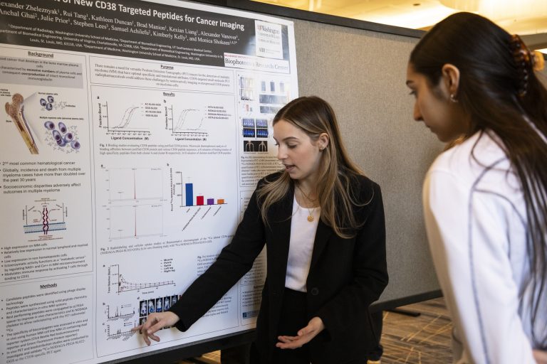
Glioblastoma (GBM) is a highly aggressive, malignant, primary brain tumor. Maximal neurosurgical tumor resection, followed by highly conformal radiation and concurrent chemotherapy, remains the standard of care for GBM patients. These tumors inevitably recur. Given the dismal prognosis, improved technologies are desperately needed to enable better treatment of recurrent GBM. An additional complicating factor is the diagnostic challenge of non-invasively discriminating recurrent GBM from radiation necrosis (RN). PARP-1, an enzyme involved in DNA repair, is selectively overexpressed in the nuclei of glioma cells.

In recent years, radiolabeled PARP-1inhibitors have been investigated for the non-invasive imaging of PARP-1 expression in glioma, as well as other tumors. Radiolabeled PARP-1 inhibitors have also been proposed for targeted radiotherapy, though, to date, there have only been a few published reports. In this project, we are examining existing radiolabeled PARP-1 inhibitors and synthesizing and evaluating novel radiolabeled PARP-1 inhibitors, as theranostic agents for GBM. Radionuclides of iodine and bromine are being used for imaging and therapeutic studies. Herein, we focus on positron-emitting radionuclides for PET imaging and Auger-emitting radionuclides for therapy. These studies are being supported by advanced MRI experiments aimed at characterizing the physiology of tumors and RN, and for assessing therapeutic response. The aims of the project are to: i) synthesize various established and novel radiolabeled PARP-1 inhibitors, and evaluate their uptake and efficacy in vitro, ii) evaluate radiolabeled PARP-1 inhibitors for uptake in murine models of GBM and experimental RN, and iii) determine the therapeutic efficacy of the PARP-1 inhibitors in murine models of GBM.

If successful, this approach will provide a new technology driven paradigm for treating patients with recurrent glioblastoma.

Imaging and Targeted Auger Radiotherapy of High-Grade Glioma

(2 of 2)

Early detection of Alzheimer’s disease (AD) remains a critical unmet clinical need. Poly(ADP-ribose) polymerase-1 (PARP-1) is a DNA repair enzyme that regulates the expression of pro-inflammatory factors. Microglia, the primary inflammatory cellsof the brain, are intimately associated with amyloid plaques in the brains of patients with AD. It has been shown that the response of microglia to neuroinflammation is mediated by PARP-1 and that PARP-1 inhibition or knockout in transgenic AD mice has a beneficial effect on cognitive dysfunction, synaptic damage, and microglial activation. To date, however, there have been no studies that have investigated a radiolabeled PARP-1 imaging agent for determining PARP-1 expression in AD mice and correlating the imaging findings with AD pathology. Building on our preceding project, which focuses on the development of radiolabeled PARP-1 inhibitors that can be used as tracers for non-invasive positron emission tomographic (PET) imaging of recurrent glioma, in this project we test the hypothesis that similar inhibitors can be used to non-invasively track neuroinflammation related to AD pathology.
We hypothesize that radiolabeled PARP-1 inhibitors will demonstrate specific uptake and retention in a mouse model of AD, compared with age-matched controls. Using in vivo PET imaging as a readout, we further hypothesize that these radiolabeled inhibitors will effectively measure changes in PARP-1 expression in cohorts of aged AD mice and that PARP-1 levels will correlate with the extent of disease progression. The aim of the project is to correlate the specific binding of a radiolabeled PARP-1 analog with Aβ plaques in the brains of AD mice. PET imaging data will be supported by, and correlated with, MR imaging results from an established pipeline of 1H MRI experiments aimed at assessing brain anatomy and physiology.

If successful, our approach will demonstrate the feasibility of non-invasive imaging of PARP-1 as an early marker of AD and provide the preliminary data for subsequent grant applications.

Our People

The lab partners with Washington University’s leading researchers in the field of magnetic resonance imagine (MRI) and magnetic resonance spectroscopy (MRS).