

Liu’s pivot was indicative of the booming growth happening within the department’s radiochemistry program. MIR was leading the charge in expanding the field’s impact well beyond isotope production and into a collaborative discipline connecting biology, chemistry, physics, medicine, computational science and more.
PET Pioneers
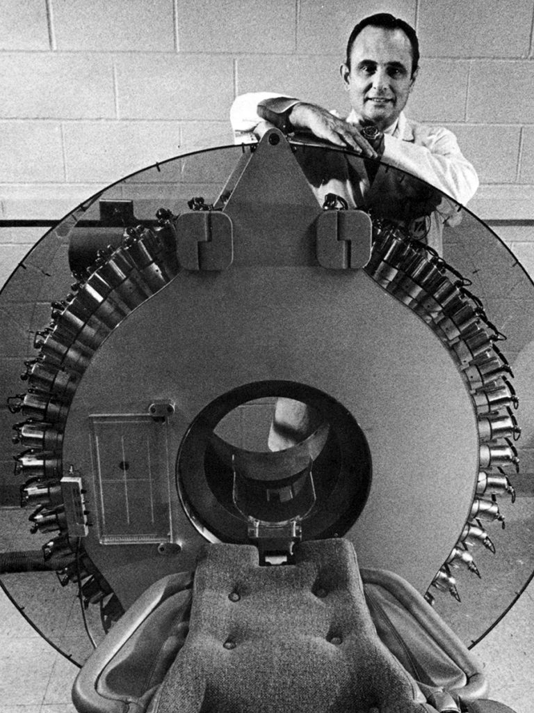
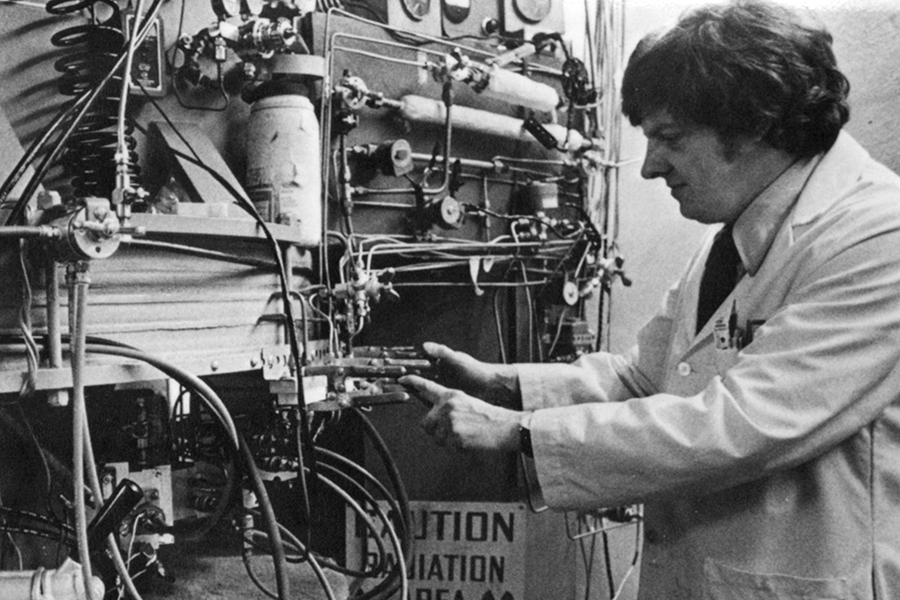
That pioneering culture didn’t emerge suddenly. It traces back to the mid-20th century, when physicist Michel Ter-Pogossian, PhD, joined MIR. Ter-Pogossian arrived in 1950, and by 1963, his team had installed the first biomedical cyclotron on a U.S. medical campus. In the 1970s, they developed the world’s first positron emission tomography (PET) scanner, famously transforming PET from theory into clinical practice. Ter-Pogossian is remembered as one of the fathers of PET, and his work established the technical base that made radiochemistry indispensable.
Welch, a mentee of Ter-Pogossian, joined MIR in 1967 and helped establish radiochemistry as a cornerstone of nuclear medicine. He emphasized what Liu recalls as the “triangle” of discovery: synthetic chemists to design molecules, radiochemists to label them and physicians to translate them into human studies.
“No matter what we do, the question is, ‘How is this going to impact human health?’”
What followed was a steady expansion: isotope production under rigorous FDA standards, the emergence of a robust radiochemistry program, and the collaborative ethos that Welch and his colleagues would bolster. When Robert J. Gropler, MD, senior vice chair and division director of radiological sciences, describes the current state of MIR’s PET research, the impact of those advancements over the past 50 years sharply comes into focus.

“We really have expertise in all the domains necessary to perform PET at a biologically specific scale,” Gropler said. “From fundamental biology to tracer development, from preclinical to clinical research, and physics support to optimize — and now computational advances that make the process more seamless and move translation even more quickly. We’re tightly integrated across the board.”
For Gropler, radiochemistry is a linchpin — the piece that gives imaging its story. Without it, PET scanners risk being cameras without context; with it, biology becomes visible in real time. “No matter what we do, the question is, ‘How is this going to impact human health?’” he said. “Linking biology to imaging, and imaging to therapies. That’s what drives precision medicine.”
From Foundation to Translation
Since the early decades of PET at MIR, the program has scaled into a national engine for radiotracer innovation. Two major NIH center grants, the Program of Excellence in Nanotechnology (PEN) and the PET Radiotracer Translation and Resource Center (PET-RTRC), were instrumental in propelling MIR into its next era.
Launched in 2005 with a five-year, $12.5 million award, PEN— led by Welch and Karen Wooley, PhD — brought together chemists, radiochemists, bioengineers, materials scientists, biologists and physicians to push nanoscale agents from concept to clinic. Wooley, currently at Texas A&M University, previously served as the James S. McDonnell Distinguished Professor in Arts & Sciences, working closely with Welch and several PEN collaborators. The effort produced one of the few nanoparticle tracers nationwide to reach human imaging. “We had to bridge the chemistry, the animal studies, the regulatory paperwork, the GMP [Good Manufacturing Practices] production. It was a complete education in how translation really works,” Liu said.
“It sounds like science fiction, but it’s already starting to happen.”
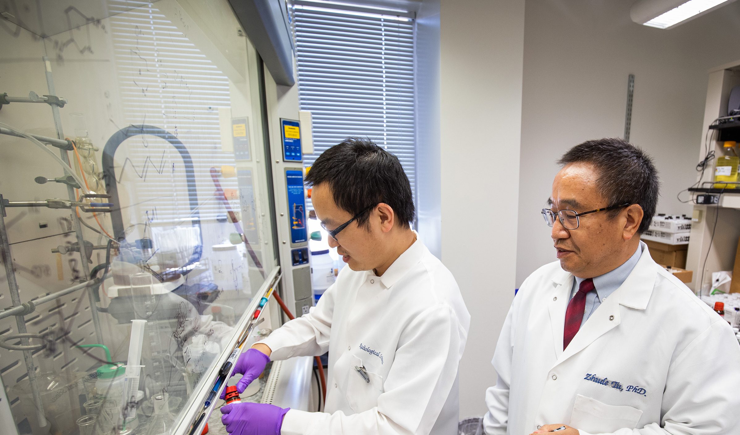
That drive toward translation continues with the PET-RTRC, a five-year, $6.3 million grant first awarded in 2018 and renewed in 2023. The PET-RTRC — led by Gropler and Zhude (Will) Tu, PhD, professor of radiology — develops and disseminates PET tracers for preclinical and human imaging studies across the country, accelerating timelines and mentoring the next generation of researchers.
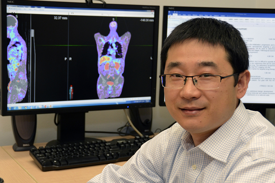
“We’re really taking advantage of our institution having such expertise in radiochemistry, in translating this science and as a GMP facility,” said Tu, who also leads MIR’s Precision Radiotheranostics Translation Center. “With this innovative technology, we’re able to drive the field forward.”
Together, these efforts positioned MIR not only to pioneer tracers, but to create the infrastructure to translate discoveries rapidly, distribute them broadly and collaborate adeptly via a push-pull mechanism with fellow researchers around the world. That vision is underpinned by an unmatched isotope supply. “We make the best copper-64 in the world,” Liu said with a smile. “Other centers wait a day for shipments; we get it straight from production. That makes an enormous difference for research.”
The Next (Not Final) Frontier
With roots in biomathematics with a focus on imaging, Kooresh Shoghi, PhD, came to MIR because imaging offered the kind of quantitative data as inputs that his mathematical models of biology needed.
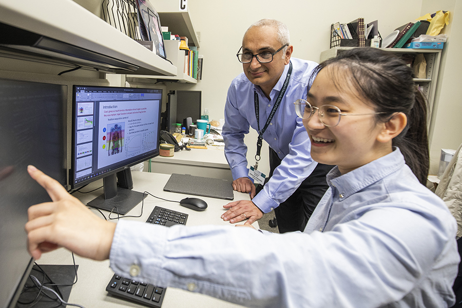
Shoghi, professor of radiology, initially set his sights on applying the tools in MIR’s radiochemistry program to develop an imaging-based systems biology paradigm to evaluate whole-body metabolism in healthy, obese and diabetic mice. His recent work includes development of quantitative co-clinical imaging trials, an innovative strategy in which patient tumor samples are implanted in mice, allowing retrospective or prospective studies with insights relevant to human disease. “Historically, researchers used tumor cell lines that had little relevance to patients,” he said. “Now, we can align quantitative preclinical and clinical imaging in ways that actually inform therapy and predict therapeutic outcomes by integrating with other multiscale data.”
Shoghi said he also looks forward to incorporating computational methods that will be central in the coming era: virtual patients and virtual imaging trials. These will be crucial to testing therapies and predicting outcomes virtually, with PET – and imaging broadly – playing a key role in informing and constraining the trials. “It sounds like science fiction,” he said, “but it’s already starting to happen.”
“Together, [computational methods and theranostics] will truly change how we practice precision medicine.”
Welch shared this vision for the future. While sorting through old file cabinets, Shoghi came across a letter dated February 2000 that revealed just how far ahead Welch had been in his thinking. In it, he urged Siteman Cancer Center leadership to hire faculty in targeted radionuclide therapy — what we now call radiotheranostics. Welch predicted the field would soon become a major clinical force. “He was early by 20 years,” Shoghi said “but he saw it coming.”
That vision is now reality. Today, MIR’s theranostics program — spearheaded by former MIR director Richard L. Wahl, MD, and currently led by Vikas Prasad, MD, PhD — has been designated a Radiopharmaceutical Therapy Center of Excellence by the Society of Nuclear Medicine and Molecular Imaging. Physician-scientists Wahl, Farrokh Dehdashti, MD, and Barry A. Siegel, MD, are a few who helped pioneer the field of theranostics, significantly expanding the use of PET in both research and clinical settings and advancing oncologic imaging through conducting some of the first human studies for novel PET radiotracers.
Gropler sees computational science and theranostics as twin frontiers defining MIR’s future. “Computational methods make translation faster and more seamless, while theranostics gives us the ability to directly link imaging to therapy,” he said. “Together, they will truly change how we practice precision medicine.”
Gropler also emphasized the need for collaboration beyond academia. MIR is already partnering with therapeutic companies to advance new imaging probes and radiopharmaceutical therapies — work that aligns academic discovery with the speed and scale of industry. “We can do the development industry can’t,” Gropler said, “and together we can bring [new therapies] to patients much faster.”
The Future at Work
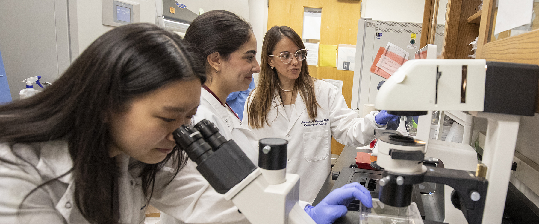
For Patrícia M. Ribeiro Pereira, PhD, assistant professor of radiology, joining MIR in 2021 was about finding the right place to build a career. Fresh from her postdoctoral training under renowned radiochemist Jason Lewis, PhD — himself a former postdoc of Welch — she was encouraged by her mentor’s experiences and by the reputation MIR carried as a center of tracer innovation. Even during her time at Memorial Sloan Kettering Cancer Center, she had worked with copper-64 that came directly from St. Louis.
“It makes the history feel real, but it also pushes you to think about what comes next.”
Ribeiro Pereira uses PET as a primary tool to tackle one of oncology’s central puzzles: why some tumors resist therapy while others respond. She is developing novel approaches to not only diagnose, but to direct treatment in a multitargeting manner capable of interrogating different tumor features simultaneously while also reversing tumor resistance. Ribeiro Pereira’s research also integrates PET with other advanced techniques to better understand how drugs distribute differently within the same tumor, teasing apart regions that resist therapy even when the drug is present.
“Tumors are not static,” she said. “They’re dynamic. With imaging, we can understand and visualize that heterogeneity in real time and in the whole tumor. That opens the door to improving existing drugs and moving closer to precision medicine.”
Her lab sits in the same space where Welch once worked. Just down the hall, his former office has been preserved as the Welch Library, lined with photographs and awards. Walking past it each day, Ribeiro Pereira feels nostalgia but, more so, motivation. “It makes the history feel real,” she said, “but it also pushes you to think about what comes next.”
Published in Focal Spot Fall/Winter 2025 Issue