Biomedical Optics and Neurovascular Imaging Lab

Projects

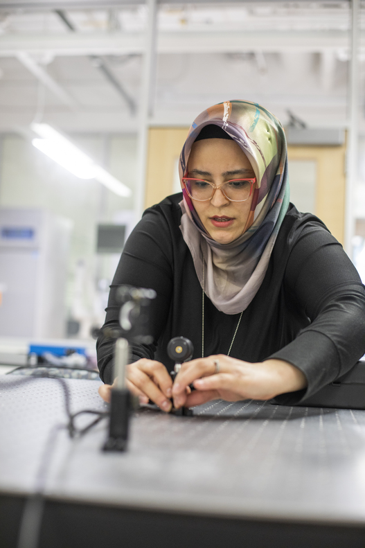
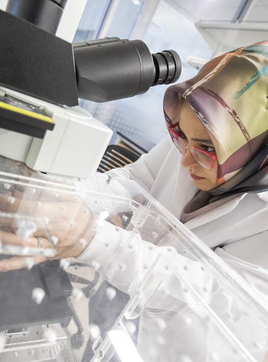
Our lab engineers advanced and accessible optical microscopy methods (such as two-photon phosphorescence lifetime imaging (2PLM), optical coherence tomography (OCT), laser speckle contrast imaging (LSCI), optical intrinsic signal imaging (OISI), and computational on-chip microscopy for understanding and improving human health through a wide range of biomedical applications.

Understanding the Brain in Action During Healthy and Diseased Conditions

The development and use of advance optical imaging methods to quantify diversity in neurovascular coupling response across physical compartments and age groups.

Healthy brain function requires abundant and uninterrupted supply of oxygen and glucose through bloodstream. The change in neuronal activity causes local vascular dilation or constriction, such that cerebral blood flow (CBF) changes accordingly to meet the metabolic demand; consequently, local changes in oxygenation (partial pressure of oxygen, pO2) follow.

This response to brain activity (a.k.a. neurovascular coupling or hemodynamic response) can show diverse characteristics across vessel types (e.g. arteries, veins, capillaries, collaterals), brain functional regions and cortical layers, over time-periods (e.g. during aging), and in abnormal physiological states (e.g. anesthesia, respiratory and/or pharmaceutical challenges, diseases). We measure the diverse characteristics of hemodynamic response in unanesthetized mice. Additionally, we investigate, how hemodynamic response differs during aging and in diseased/challenged conditions.

  • National Institutes of Health (BRAIN Initiative R00MH120053)
  • Mallinckrodt Institute of Radiology
  • McDonnell Center for Systems Neuroscience
  • McDonnell Center Cellular and Molecular Neurobiology

Quantitative Interpretation of BOLD fMRI Signal and Bridging Knowledge Across Imaging Modalities

Optical measurements of oxygenation and blood flow in awake mouse with high spatial and temporal resolution during standard fMRI calibrations (hyperoxia, hypercapnia, caffeine intake etc.).

Comprehensive bottom-up computational models of BOLD signal are using realistic vascular anatomical network (VAN) simulations of the fMRI voxel at the capillary scale. These computational models are constructed on data collected from well-designed experiments in animals, providing independent optical measurements of partial pressure of oxygen (pO2), cerebral blood flow and vascular structure. By achieving a more realistic and unrestricted insight into the neurovascular coupling, they have been indispensable tools in a quest to develop quantitative BOLD fMRI. However, these models are still limited by the experimental conditions (anesthetized animals, normal breathing conditions, young age, limited cortical depth, etc.) in which data is obtained.

To address this need, we investigate effects of mild respiratory and pharmaceutical challenges (commonly used for BOLD fMRI calibration) on the hemodynamic response in awake animals, across cortical layers and in different functional regions. Our research aims to provide a ‘textbook’ description of cerebral microvascular oxygen delivery, to inform physiologically realistic bottom-up modeling efforts of BOLD signal, and to enable future studies of neurovascular coupling and the role of vascular dysfunction in neurodegenerative diseases.

Identifying Early Markers of Neurovascular Diseases and Preventative Strategies

Quantifying changes in retinal and intestinal oxygenation, flow and microvascular structure during disease progress, their correlation with brain, and relationships with sleep, diet and exercise.

The eye and gut are two organs whose abnormalities have been strongly linked to the pathophysiology of various neurodegenerative diseases. For instance, several retinal microvascular changes have been associated with Alzheimer’s disease and are expected to predict the disease progression in brain. Additionally, studies on the microbiota-gut-brain axis reveal that imbalances (dysbiosis) in gut microbiota diversity are linked to several neurological disorders, including stroke, Alzheimer’s and Parkinson’s diseases. Impaired oxygen delivery in the intestines is one among many factors that may lead to dysbiosis of gut microbiota.

Recently, we have transferred our expertise in two-photon microscopy for brain imaging into the measurement of oxygen metabolism of eye and gut. In mouse retina, we investigate oxygenation in the three-layer capillary plexus, non-invasively through the pupil under different breathing conditions. Also, we investigate the oxygenation and red blood cell flux in the capillary bed of the healthy mouse intestine. We plan to use these methods for quantification of (1) changes in retinal/intestinal oxygenation during disease progress, (2) their correlation with changes in cerebral oxygenation, and (3) their relationship with sleep, diet, and exercise.

More in Biomedical Optics and Neurovascular Imaging Lab

Latest in the BRC