Breast Imaging FAQ
Jump to:
Screening
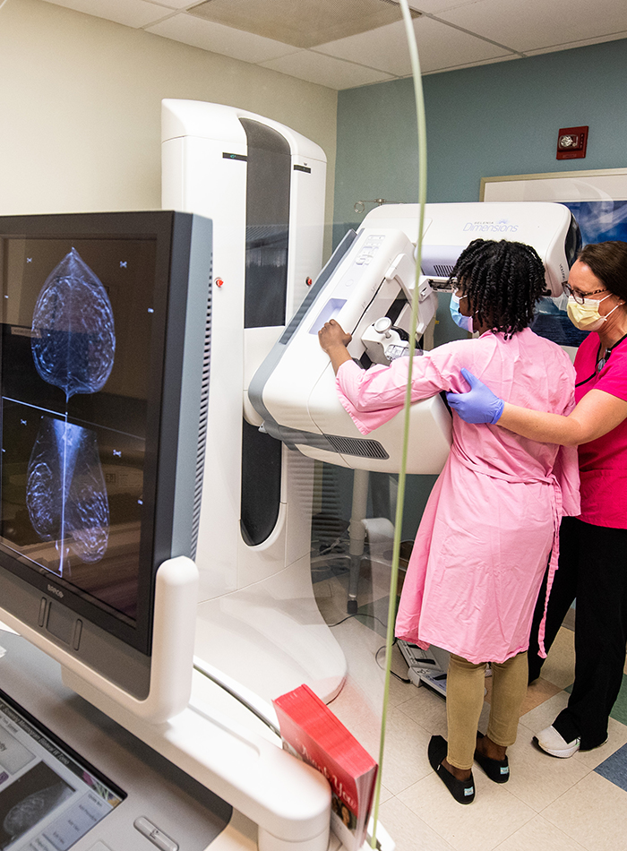
Screening mammograms are recommended annually for all women beginning at age 40. However, if there is a history of breast cancer in your family, your doctor may assess your risk factors and recommend screening before age 40.
3D mammography is an advanced type of non-invasive, low dose imaging that combines multiple high-resolution breast X-rays to create a three-dimensional picture of the breast. It provides images of the breast in “slices” from several different angles, making some abnormalities easier to see. This aids in the early detection and diagnosis of breast cancer. Unlike some other imaging centers, MIR uses 3D mammography in all imaging facilities, including our mammography vans.
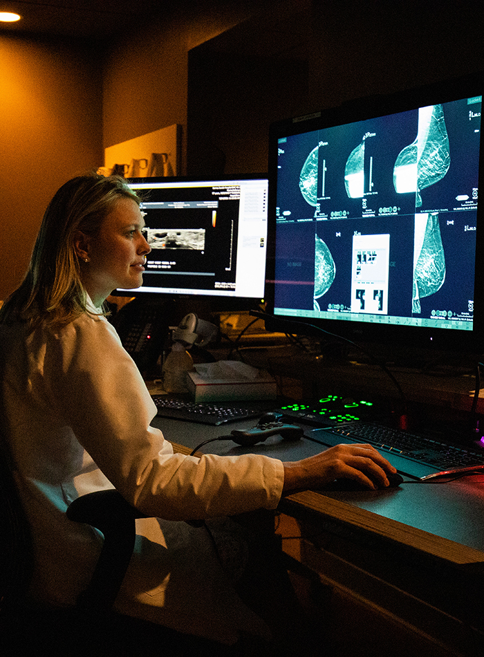
Getting called back after a screening mammogram is fairly common. It means that the doctors have found something they want to look at more closely with additional images. Fewer than 1 in 10 women called back for more images are found to have breast cancer.
You can ask the imaging center to load any prior mammograms onto a CD, then bring the CD with you on the date of your exam so an MIR radiologist can compare your past mammograms with your new images.
Dense breast tissue is a normal and common finding. When viewed on a mammogram, women with dense breasts have more dense tissue than fatty tissue. Because dense tissue increases the chance that breast cancer may go undetected by a mammogram, your doctor may recommend an additional screening exam, such as breast ultrasound or breast MRI.
Diagnostic
A diagnostic mammogram is ordered by a physician when further evaluation of the breast is needed. It is often used to investigate suspicious breast changes, such as a new breast lump, breast pain, an unusual skin appearance, nipple thickening or nipple discharge. It is also used to evaluate abnormal findings on a screening mammogram. A diagnostic mammogram usually involves getting additional images of the breast and may include breast ultrasound or breast MRI, depending on your risk factors.
Breast ultrasound technology uses sound waves to evaluate specific areas of concern that can be felt, such as a breast lump, or anything identified on a mammogram that requires further study. It is often used in combination with diagnostic mammography. While mammography images evaluate the entire breast, ultrasound images can provide more information on a specific area.
Breast MRI is a highly advanced technology that uses magnets and radio waves to produce images of soft tissue in the breast. When used with screening and diagnostic mammography, MRI technology provides additional information for the early detection and diagnosis of breast disease. Breast MRI can also serve as an additional safeguard for women with a high risk of breast cancer.
Biopsy
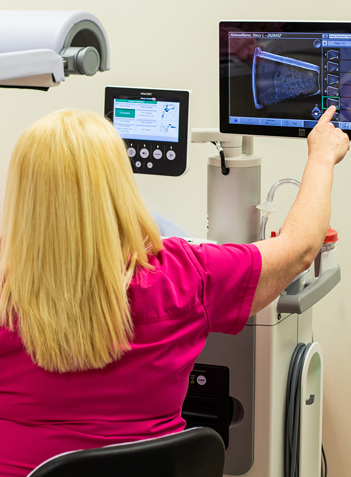
A biopsy is a test that removes tissue or fluid from the breast to be looked at under a microscope for further evaluation. MIR radiologists regularly perform three different kinds of biopsies: fine needle aspiration, image-guided core needle biopsy and a surgical biopsy. Learn more.
A very thin needle is used to draw cells from the area of the breast that has suspicious changes in order to provide a diagnosis without surgical intervention. There is minor discomfort with this procedure, which typically lasts just a few seconds.
An image-guided breast biopsy uses a hollow needle to remove a small sample of breast tissue or cells for examination under a microscope. MIR radiologists regularly performs three types of image-guided biopsies:
- Stereotactic biopsy
- Ultrasound-guided biopsy
- MRI-guided biopsy
A surgical biopsy is a brief procedure that requires light sedation but not an overnight stay. A small incision is made to remove either the entire mass of suspicious breast tissue or a representative sample, depending on its size and location.