

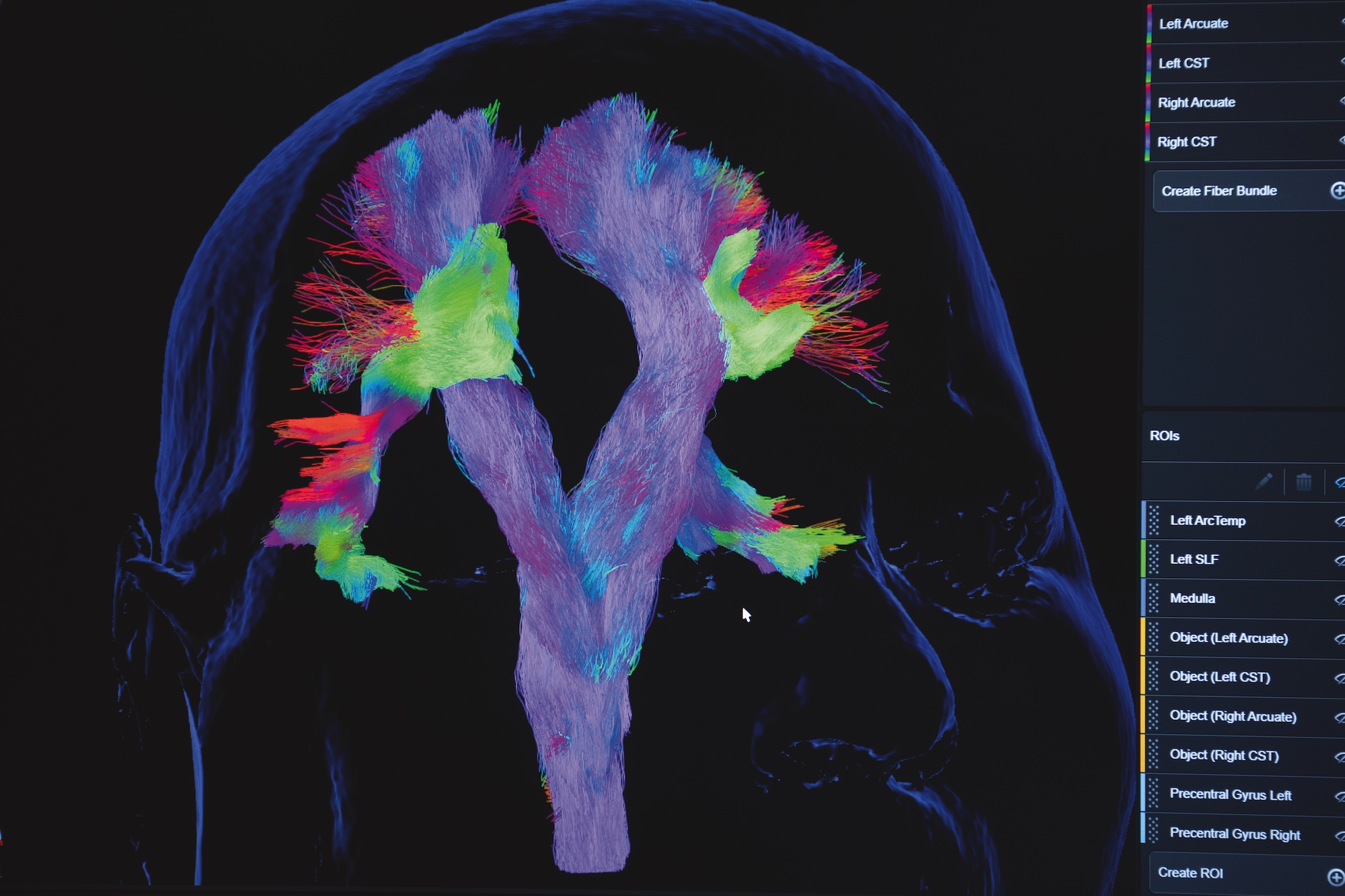
AI-powered software designed to rapidly chart the brain’s functional landscape — pinpointing regions involved in speech, vision, movement and other crucial abilities — has been authorized by the Food and Drug Administration (FDA). The program can now be offered to hospitals to support precision neurosurgery, from treating epilepsy to performing intricate surgeries and removing brain tumors.
Behind a milestone as significant as FDA approval are years of cross-disciplinary work. In this case, it included a long arc of discovery from WashU Medicine Mallinckrodt Institute of Radiology (MIR) physicians and scientists. MIR’s contributions in the areas of functional imaging of the brain, data infrastructure and clinical workflows played a key role. Tammie L.S. Benzinger, MD, PhD, the Hugh Monroe Wilson Professor of Radiology, and Daniel S. Marcus, PhD, professor of radiology, unpack the advanced imaging and informatics that helped empower the successful translation of this project.
Let’s rewind a bit. How does this milestone connect to WashU’s longer history in resting-state functional magnetic resonance imaging (fMRI)?
Marcus: There’s a direct throughline from the transformative foundational work of Marcus E. Raichle, MD — who played a central role in the understanding of a “default mode network” to describe resting-state brain function — to what we’re doing today with AI and neurosurgical mapping.
Benzinger: When this project started, we had a really large data set of healthy adult resting-state brain MRIs collected through studies of aging at the Knight Alzheimer Disease Research Center (Knight ADRC) at WashU. Within the Neuroimaging Labs Research Center (NIL-RC) are pioneers of fMRI, including Abraham Z. Snyder, MD, PhD, and Joshua S. Shimony, MD, PhD, who are co-investigators with neurosurgeon Eric Leuthardt, MD, on this clinical translation of brain mapping technology. Avi and Josh were pivotal to moving resting-state science forward, as were radiologists Michelle M. Miller-Thomas, MD, and Gloria J. Guzmán Pérez-Carrillo, MD, who advanced the clinical workflows. Few places have this combination of continuity of knowledge and the blend of neuroscientists, physicians and informatics engineers needed to translate it into real-world clinical practice.
FDA clearance can take years. What MIR-based expertise helped move this project forward?
Marcus: Numerous MIR researchers across multiple research centers contributed to the algorithm development work, which combined radiology, neurology, neurosurgery and computational imaging expertise to build a machine-learning model that can identify functional brain areas from patient data. On the infrastructure side, the group I previously led (the Computational Imaging Research Center) and 1Rad (MIR’s IT support) helped build the data platforms and workflows to pull exams from hospital systems, run processing reliably and return results into clinical systems. We’ve had to develop platforms and an ethos that is centered around translation and corresponding efficiently with hospital systems. It’s a big feat.
What were some of the make-or-break technical challenges of this project?
Marcus: We needed perfect spatial registration. These maps ultimately guide surgery, so the images and network maps have to line up precisely — from what’s collected on the scanner all the way to what the surgeon sees on the navigation system in the operating room. We spent a lot of time validating registration end-to-end, making sure it worked everywhere, because it had to be perfect.
Benzinger: This only works if you can collect the right data consistently and with high quality. When scanners arrive from the factory, they’re not optimized for the clinical questions we need to answer. One of MIR’s strengths is tailoring protocols. We build exams that match the patient and the decision at hand and enforce quality control so the data from the clinical scanners produce data comparable to our research scanners, for example.
In the relay race from discovery to clinical impact, collaboration makes a huge difference. What made that cross-disciplinary handoff work here?
Benzinger: The culture. We work in a really integrated fashion. This work brought scientists, radiologists and neurosurgeons together around patient care, not credit. The lack of “walls” meant we could move unusually fast: from the project’s origins with then-MD/PhD student Carl Hacker’s research analysis all the way to a clinical service now led by Matt Glasser, MD, PhD — which is used for essentially every neurosurgical brain tumor patient at Barnes-Jewish Hospital.
What’s next and how will this scale beyond WashU?
Marcus: With AI advancing, I think we’ll see more precise maps, larger datasets and new disease areas. Very likely there will be impactful clinical applications in psychiatric disorders, neurological disorders and dementia. Since we started this project years ago, there have also been major leaps in the efficiency with which WashU innovators are able to commercialize their work. Combine that with the new AI-focused research center at MIR, and we’ll be well positioned to develop and scale projects like this one and beyond.
Published in Focal Spot Spring/Summer 2026 Issue