
AI applications are rich across the Mallinckrodt Institute of Radiology (MIR) research enterprise. Aimilia Gastounioti, PhD, is utilizing AI to assess long-term breast cancer risk and address racial disparities in breast cancer screening. Aristeidis Sotiras, PhD, is characterizing Alzheimer’s disease heterogeneity and working to improve follow-up for patients with incidental findings. Abhinav Jha, PhD, develops and rigorously assesses AI algorithms to determine their viability for enhancing, reconstructing and evaluating medical imaging. Debbie L. Bennett, MD, is evaluating the clinical impact of computer-assisted breast cancer detection. And Vamsi R. Narra, MD, has helped scale AI applications across MIR’s clinical operations, from computed tomography (CT) to magnetic resonance imaging (MRI).
“With AI, it applies to everything and everywhere,” said Robert J. Gropler, MD, professor of radiology and senior vice chair and division director of radiological sciences. “We’ve invested heavily in the intellectual expertise — our faculty — and now we’re doubling down on the infrastructure piece.”
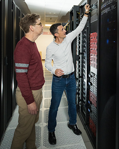
Enter the Research Computing and Informatics Facility (RCIF).
There’s a New Facility in Town
The RCIF provides a breadth of services covering hardware, software, training, support and data. More specifically, the new facility provides users with access to a high-performance computing cluster, high-speed and bulk storage, robust systems for management of imaging and related project data, access to a deep well of anonymized clinical data for research purposes, and training sessions and workshops. All in all, the RCIF has the resources and expertise to support any computationally intense research project, according to co-directors Mike R. Hodge and Scott M. Johnson, PhD.
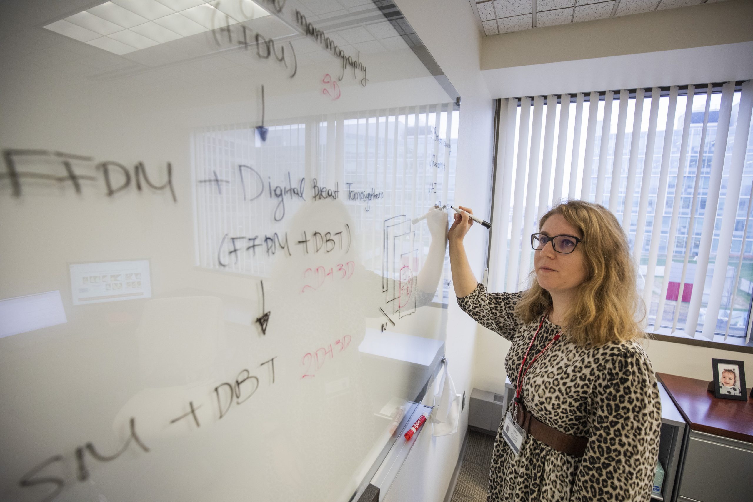
For example, Gastounioti, an assistant professor of radiology and investigator in MIR’s Computational Imaging Research Center (CIRC), is focused on a variety of mammography projects. Until now, research capabilities were often limited to 2.5D at most. But the RCIF enables computational analysis of 3D mammograms, with AI algorithms able to consider the full structure rather than settling for quasi-3D data. “Simply put, it enables bigger science,” said Johnson.
“With AI, it applies to everything and everywhere.”
Sotiras, an assistant professor of radiology, previously ran into similar roadblocks when it came to capacity. Also a CIRC investigator, he develops supervised and semi-supervised machine learning algorithms to better understand how Alzheimer’s disease (AD) pathology affects certain regions in the brain. The work he is doing to summarize high-dimensional data — often multimodal scans that show anatomy, structure and pathology in the brain — could have potential applications in diagnosing earlier and with higher accuracy which AD patients will decline and which will not.
“Ultimately, the more resources you have, the more ambitious you become with your models,” said Sotiras. “Some of our decisions were dictated by what hardware we had access to. Previously, if our models were too big, we would design smaller models to circumvent some of our resource challenges.”
Beyond expanded processing capacity — fivefold to be exact — the RCIF’s offerings will save researchers valuable time tackling thorny projects. “For a lot of people, they can get 90% of what they need to get done pretty quickly,” said Johnson. “But it’s that 10% that is hard stuff to crunch through. And we can get rid of that and get them to a useful product in record speed.”
In addition to multiple direct support channels, the RCIF offers monthly introductory training sessions — even for those with little to no high-performance computing experience.
Data is King
AI applications are endless but ultimately powerless unless researchers have access to ample data to advance their work. Fortunately, data isn’t a problem at MIR.
Building on a legacy of innovation in the computational space, the RCIF will leverage the thousands of studies available through the revolutionary Central Neuroimaging Data Archive (CNDA) and the anonymized clinical imaging sessions available through Flywheel (a biomedical research data platform). Both CNDA and Flywheel are built on XNAT, a widely used open-source imaging informatics platform developed at MIR by Daniel S. Marcus, PhD, professor of radiology and director of the CIRC.
In addition to both in-house and multisite studies, researchers have access to data gleaned from decades of collaboration with BJC HealthCare and Siteman Cancer Center. Gastounioti’s work to reduce racial disparities in breast cancer screening emphasizes the importance of not only an abundance of data, but also diversity of data. “Ultimately, we want to develop models that generalize well across racial subgroups, so we need access to diverse patient data,” she said. For example, breast density in Black women is sometimes downgraded despite being an important risk factor when it comes to cancer risk assessment, scheduling supplemental imaging and planning treatment. “In some cases, preventive interventions or supplemental imaging may not be eligible for insurance coverage if a woman doesn’t fit into a high-risk density category.”
Sotiras is also working to keep patients from falling through the cracks of the health-care system. Using existing radiology reports and AI, he aims to enhance MIR’s incidental findings program to identify whether a report should be flagged for follow-up.
“In our clinical work at MIR, we have very good resources including nurse coordinators doing follow-ups,” said Sotiras. “But imagine now that this could translate somewhere where they don’t have enough radiologists or coordinators.”
“Imagine now that this [technology] could translate somewhere where they don’t have enough radiologists or coordinators.”
In other efforts to streamline workflows and advance imaging science, Jha, assistant professor of radiology, has a vast portfolio of AI research that includes rigorous evaluation of AI algorithms and how they perform on clinical tasks. When his team evaluated a common approach to denoise cardiac SPECT images, they found the denoised and original images to be visually similar. But ultimately the denoising technique either had no significant impact or even degraded performance when it came to detecting heart defects.
Jha, also an assistant professor of engineering, said in an article from the McKelvey School of Engineering, “This emphasizes the important need for performing evaluation of AI algorithms on clinical tasks and not just relying on visual similarity as a measure of performance.”
Making a Clinical Impact
AI applications exist not only within the research space but are readily used every day in clinical practice. “There are AI applications available in the market for multiple modalities,” said Narra, professor of radiology and vice chair for clinical imaging informatics and new business development. “We currently are piloting three of them in our clinical environment.”
“We want to educate our faculty about what’s possible now and how we can do it.”
The key element, and perhaps the trickiest, of effectively integrating AI into clinical practice is connectivity between systems. Previously, there were 11 different PACS (picture archiving and communication system that is used to review and interpret radiology images) with no way to integrate AI into the workflow. Now with a single enterprise radiology PACS, it is easier to deploy AI applications across multiple sites.
MIR has been the first in North America to utilize the Sectra AI Amplifier Service — an all-in-one service streamlining the selection, activation and use of AI applications on the enterprise PACS — which is designed to support homegrown AI applications as well.
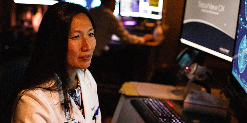
Bennett, associate professor of radiology and chief of breast imaging, has been studying several breast cancer AI applications in hopes of improving cancer detection and recall rates through the technology’s improved specificity and sensitivity. “We’re excited to incorporate AI into clinical practice to help us find cancers as early as possible,” Bennett said.
While there are a myriad of examples of AI projects already making waves across the WashU Medicine campus, “one of the most important aspects of our AI infrastructure expansion is education,” said Gropler. “We want to educate our faculty about what’s possible now and how we can do it.”
Gropler highlights an integrative approach that includes: 1. clinicians and/or basic researchers 2. computational scientists 3. entrepreneurial tech transfer resources 4. commercialization and democratization and 5. regulation. These components fuel the full lifecycle of an effective AI application that can be incorporated into clinical workflows. “Considering all of these parts is how we make a major impact.”
Published in Focal Spot Winter 2024 Issue