NeuroPhoto Lab

Projects

High-Density Diffuse Optical Tomography

Goal

To provide a more naturalistic imaging system, but address imaging quality issues with traditional fNIRS, we developed high-density diffuse optical tomography (HD-DOT) system complete with lightweight fiber optics. HD-DOT is an emerging method that uses a dense array of overlapping light measurements to improve image quality over traditional fNIRS as validated against fMRI in adults. The naturalistic imaging environment was combined with more naturalistic imaging tasks, specifically movie viewing of traditional entertainment movies. Features across these movies, such as faces and speech, can be tracked and correlated with brain activity to visualize responses similar to standard functional imaging tasks. Using these techniques, we can map brain function in a variety of subject populations that are not amenable to fMRI, including children, subjects with cochlear implants or deep brain stimulations, or using portable systems we can image people in clinical environments

Functional neuroimaging in humans has revolutionized cognitive neuroscience. Increasingly, functional neuroimaging is being used as a diagnostic and prognostic tool in the clinical setting and to view brain development. Its expanding application in the study of disease and development necessitates new, more flexible tools. The logistics of traditional functional brain scanners (e.g., MRI) are not well-suited to subjects who are in an intensive care unit or to subjects who might otherwise require sedation for imaging, such as infants and young children. Furthermore, traditional functional neuroimaging methods use behavioral paradigms that are not appropriate for these same subjects. Young children cannot reliably attend to many tasks, nor can unconscious patients in the operating room or intensive care. Functional near-infrared spectroscopy (fNIRS) is more child-friendly with its open scanning environment but offers poor image quality. Task engagement is also a challenge for children as it can be difficult to remain focused during tasks such as watching checkerboards. Naturalistic stimuli are becoming increasingly popular in cognitive neuroscience research as they are more engaging, but their complexity requires alternative data processing methods. Development of a child-friendly imaging system validated against fMRI with an age-appropriate stimuli would allow greater insight into early childhood brain development.

Mapping Multi-Scale Networks in Mice

Mapping spontaneous brain activity with functional magnetic resonance imaging (fMRI) and functional connectivity (FC) techniques has become a dominant neuroimaging assay for humans. FC mappings have implications for both basic neuroscience and clinical disease where fcMRI can map functional reorganization, neural plasticity and neural network integrity. As functional connectivity methods advance, a gap continues to grow between human fcMRI and the detailed genetic and molecular assays common in mouse models. In particular, the molecular and cellular underpinnings of brain-wide correlated brain activity are relatively unresolved, and an understanding of them will grow in importance as FC measures extend further into studies of disease.

To address this, we recently developed hemodynamic and calcium (26)  mapping of FC in mice using widefield optical imaging (WOI). WOI is sensitive to several neurological diseases (e.g. stroke and Alzheimer’s, and we have begun to unravel molecular mechanisms involved in plasticity of functional connections. While hemodynamics are label-free, genetically encoded calcium indicators (GECI’s) enable imaging in specific cell types (e.g. neurons  or astrocytes) and are a faster report of neural activity. However, a critical limitation of all WOI methods remains resolution (>100 microns) and lack of depth sectioning.

Calcium dynamics of mice with GECI have also been measured with subcellular resolution using video-rate (30 Hz) two-photon microscopy (TPM), providing an exquisite window into small scale neuronal dynamics. These TPM studies have generally focused on small fields-of-view (FOV), <1 mm, and thus are not suited to image the combination of short range and long range connections mapped with WOI. Very recently new TPM designs have begun demonstrating larger FOV’s with diameters up to 8 mm. However, these large field-of-view (LF)-TPM systems have run into the daunting challenge of speed, with full-frames rates dropping proportional to FOV area or worse. The curvature of the mouse brain poses a further challenge for LF-TPM of long range connections, because the axial sectioning is much smaller (e.g. ~10µm) than the curvature of the mouse head (e.g. axial deflections >1mm for FOV >8mm).

With these issues combined for FOV>8 mm, existing LF-TPM designs have full-frame rates <0.01Hz, and thus >1000x too slow to image long range calcium FC. The goal of this project is to develop a new fast LF-TPM (FLF-TPM) system, using a number of approaches that in combination will allow >10 Hz full-frame rates. To connect FLF-TPM with WOI results, we will integrate WOI into the FLF-TPM, develop methods for mapping FC across multiple spatial scales, and evaluate the system in studies of brain plasticity.

The goal of this project is to develop a new fast LF-TPM (FLF-TPM) system, using a number of approaches that in combination will allow >10 Hz full-frame rates. To connect FLF-TPM with WOI results, we will integrate WOI into the FLF-TPM, develop methods for mapping FC across multiple spatial scales, and evaluate the system in studies of brain plasticity.

NIH-NINDS – R01NS099429

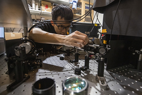
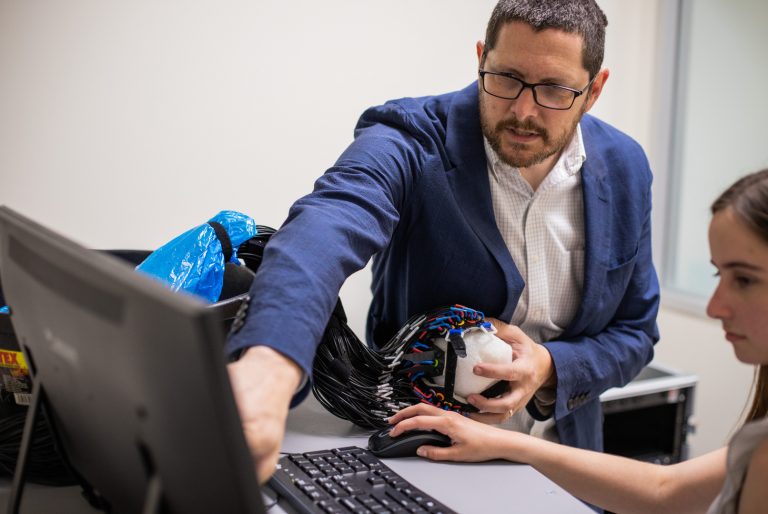
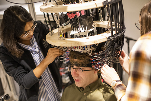
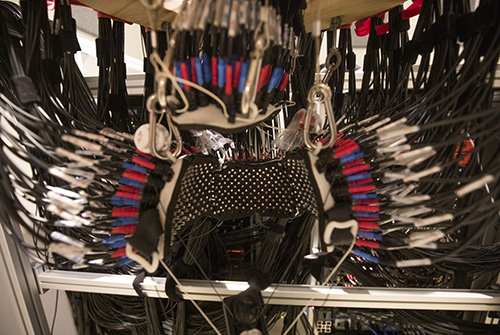
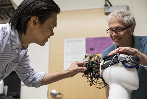
Wearable High-Density Diffuse Optical Tomography

Brain imaging with MRI machines provides noninvasive access to the neural basis of development, degeneration, and disease of the brain. However, the logistics of MRI are ill-suited to many applications. Functional MRI (fMRI) is not suited for imaging subjects who cannot lie sufficiently still in MRI scanners (e.g., awake children under 5 years old) and children with disorders of voluntary movement, such as moderate to severe cerebral palsy (CP), which represents 0.2% of infants, or 10,000 annually. Many patients with implants (cochlear implants, deep brain stimulators, heart pacemakers) are also contra-indcated for MRI.

A promising potential surrogate to fMRI is high-density diffuse optical tomography (HD-DOT) a tomographic version of functional near infrared spectroscopy (fNIRS). However, despite these advances, application of HD-DOT to naturalistic studies in children has been limited by a requirement of large opto-electronic consoles and bulky fiber optics. Several wearable fiber-less fNIRS instruments are now available commercially, but traditionally these systems have had multiple deficits including either lower resolution, lower field-of-view, strong image distortions, or less signal to noise than our fiber-based HD-DOT systems. In this project we are developing a full head WHD-DOT device that matches performance of fiber-based HD-DOT for mapping brain function in typical and atypical child development.

NIH-NIBIB, U01 EB027005

Speckle Contrast Optical Tomography for Imaging of Cerebral Blood Flow

Goal

Real-time maps of cerebral blood flow (CBF) at the bedside are a long sought-after assay for neurocritical care. Regional CBF measures can indicate which brain regions may be becoming ischemic and are at danger for hypoxic-ischemic injury. The dominant clinical methods for mapping microvascular CBF include PET and arterial spin labeling (ASL) with MRI. However, PET and ASL-MRI provide only snapshots of CBF, not continuous monitoring, and thus miss dynamic events. Cerebral blood flow dynamics are important in many clinical scenarios, including acute stroke, traumatic brain injury and preterm birth. To monitor brain perfusion or its surrogates at the bedside clinically, several non-imaging methods are used. These include intracranial pressure (ICP), a thermal dilution method, and laser Doppler flowmetry, all of which are either very invasive or only very local measures. Unfortunately, none of these existing monitoring methods provide the desired regional map of CBF at the bedside.

Noninvasive optical techniques are an attractive approach for measuring brain blood dynamics at the bedside and address a gap in current clinical CBF imaging. We are developing speckle contrast optical tomography (SCOT), a new method for transcranial optical imaging of CBF in humans. Our approach aims to adapt laser-speckle-based methods for estimating CBF with high-density measurement arrays to facilitate tomographic imaging. To enable cost-feasible instrumentation, we are developing speckle contrast methods using low-cost multi-mode optical fibers that can be implemented in parallel in modern scientific CMOS cameras to achieve sufficient channel count for high-density SCOT imaging. To image with SCOT data, we will develop algorithms that model diffusion of speckle contrast measures in anatomically realistic head models then invert those models to build images of CBF. This tomographic synthesis of the data allows separation of brain signals from scalp and skull artifacts. Our high-density SCOT systems will be evaluated in comparison to MRI and PET before establishing feasibility of imaging CBF in ischemic stroke patients.

NIH R01NS090874

Our People

The NeuroPhoto Lab, led by Joseph Culver, PhD, leverages multidisciplinary excellence in the areas of electrical engineering, biomedical engineering, computational imaging, physics and neuroscience.

Latest in the BRC