New MRI Scan Studies Overlooked Brain Cell Data

MRI scans generate a wealth of data, but most researchers are only interested in a small portion of the signal and ignore the rest of it.
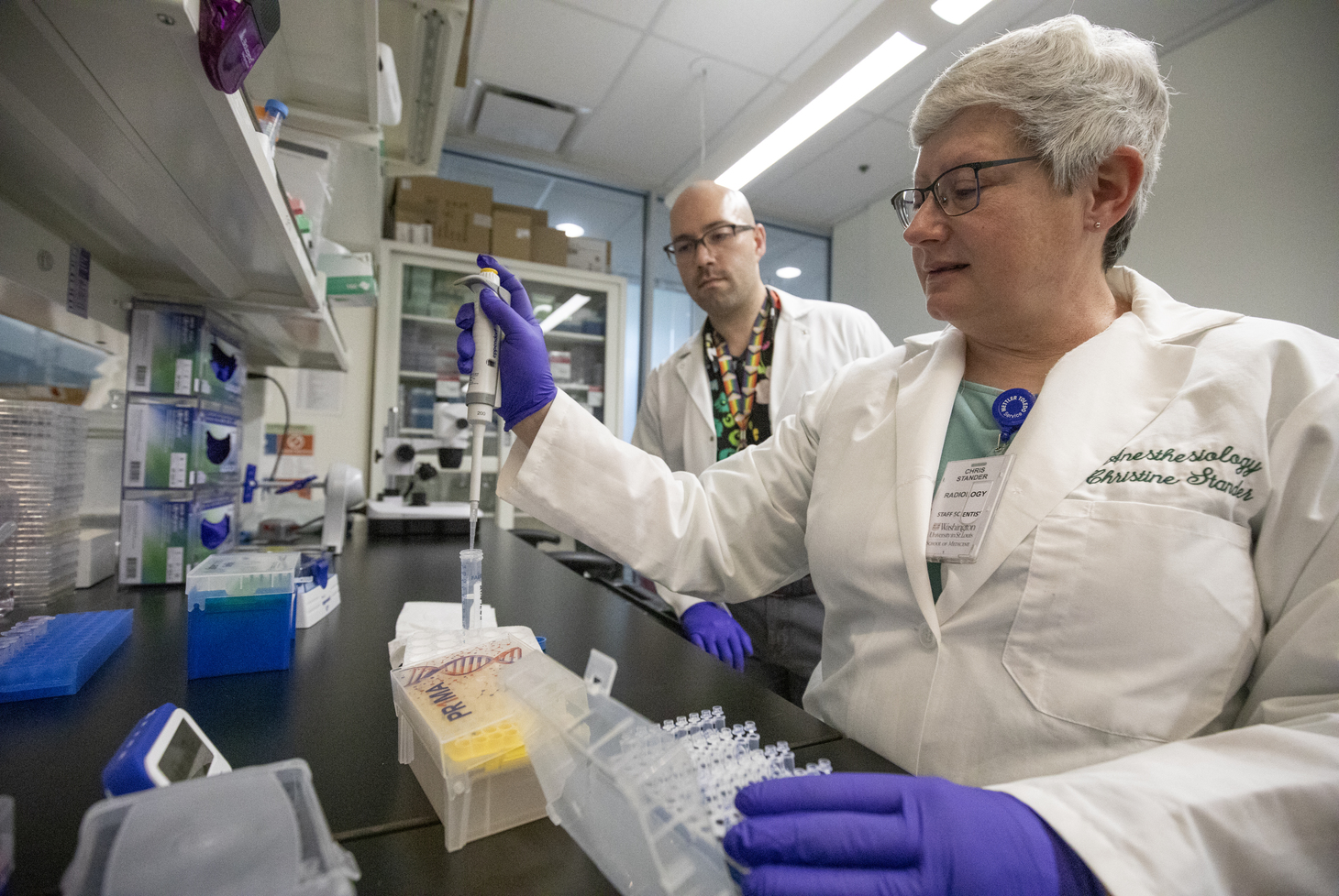
Dmitriy A. Yablonskiy, PhD, a professor of radiology for Mallinckrodt Institute of Radiology at Washington University School of Medicine, directed a study with Marcus E. Raichle, MD, the Alan A. and Edith L. Wolff Distinguished Professor of Medicine, where they analyzed MRI background data to reveal how many and which brain cells are present in the scan and show where cells have been lost through injury or disease.
The MRI tracks a signal, which they called R2t*, and only takes six minutes to complete. This data gave the researchers all they needed to know to determine how densely packed and interconnected the neurons are in any part of the brain. Their findings, published in Proceedings of the National Academy of Sciences, may eventually help us understand how a person’s brain develops or give clues to diagnosing illness or injury through a simple scan.
Read more on the School of Medicine’s website.