

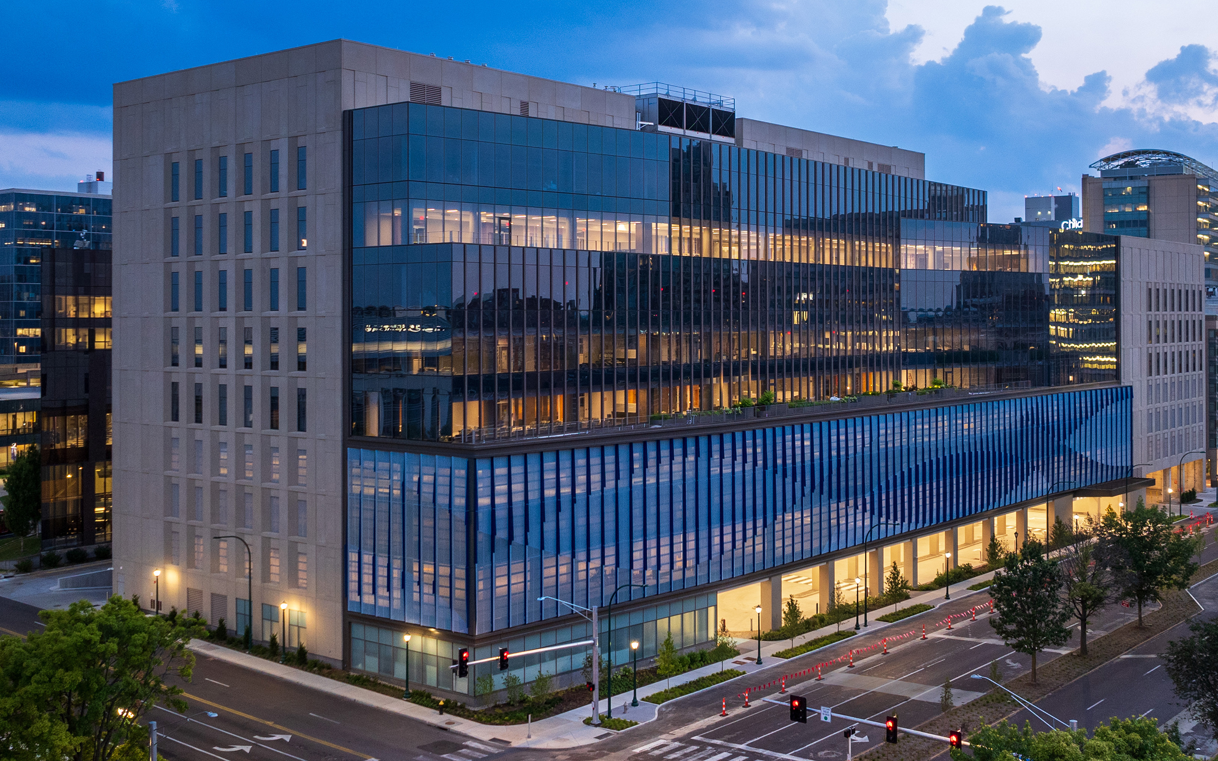
The vision behind the new Gary C. Werths Building at Siteman Cancer Center seeks to center the patient and lift some of the burden of care from their shoulders, creating a streamlined and personalized experience that includes state-of-the art imaging services from WashU Medicine Mallinckrodt Institute of Radiology (MIR).
Patients visiting the Werths Building for outpatient care will receive a coordinated, single visit. They may first see a multidisciplinary team of WashU Medicine cancer experts, then receive chemotherapy and other services in collaboration with BJC HealthCare caregivers. And when needed, patients may undergo imaging studies provided by MIR radiologists.
Leading-Edge Modalities in the House
In addition to 3 Tesla MRI and ultrasound, the building’s portfolio includes a whole-body PET/CT and photon-counting CT. The latter, a Naeotom Alpha® photon-counting CT scanner, was approved by the Food and Drug Administration in 2021. At the time, it was described as the first major computed tomography advancement in nearly a decade. Unlike other CT scanner detectors, which measure the total energy contained in many X-rays at once, the Naeotom Alpha CT’s detectors measure each X-ray passing through a patient’s body. The information is converted into a detailed, 3D image that can be used by MIR radiologists to help diagnose or prepare a plan of treatment for patients.
“This state-of-the-art photon-counting CT allows us to push the envelope on lowering radiation dose, scanning patients more quickly and deriving more information from CT scans than earlier generations of scanners,” said Vincent M. Mellnick, MD, chief of abdominal imaging and director of radiology services at the Werths Building.
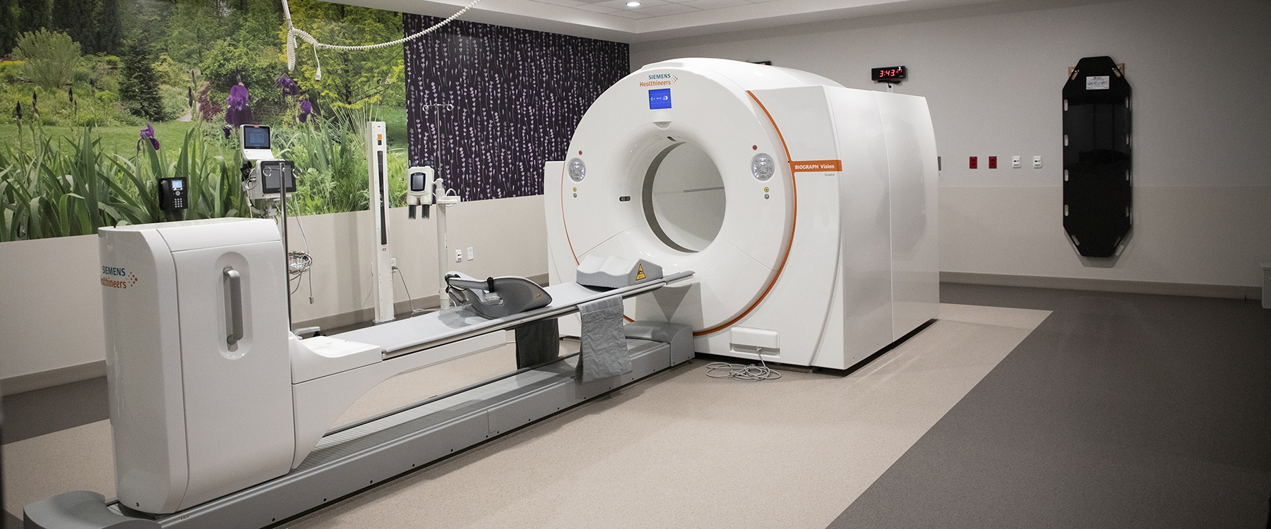
The new outpatient cancer care center is also home to a Biograph Vision Quadra™ PET/CT scanner. MIR is among the first radiology departments in the country to install this advanced diagnostic imaging scanner. “This scanner enables us to perform whole-body imaging in only a few minutes,” said Farrokh Dehdashti, MD, senior vice chair and division director of nuclear medicine. Similar to the photon-counting CT at Werths, the high sensitivity of this advanced technology enables shorter exam duration and lower radiotracer doses, minimizing patients’ radiation exposure while maintaining excellent image quality.
“Both the shorter scan time and reduced radioactivity are extremely important for our oncology patients, who often are not feeling well and cannot lie down for a long period of time,” added Dehdashti. With its ability to scan the whole body at once, the scanner also has potential for use in research studies in areas such as metabolism and perfusion.
An Embedded Reading Room
Within the neurosurgery clinic in the Werths Building exists a feature that may be one of its kind — an embedded neuroradiology reading room. Imaging plays a central role in managing patients with brain tumors. But typically, neuroradiology reading rooms are located in areas separate from neurosurgery clinics, such as in a radiology department. “We saw this new neurosurgical clinic as an opportunity to be in close proximity to our surgeons and oncologists so that they can immediately consult with us should they have questions regarding a patient’s images,” said Katie D. Vo, MD, professor of radiology and chief of neuroradiology.

Conveniently, today’s technology enables radiologists to read imaging studies remotely from across the globe and while in their offices or homes. But those capabilities, Vo noted, can distance the human connection between radiologists and other providers.
“This ambulatory cancer center is designed with a philosophy of centering the patient and collaboration among medical professionals,” said Vo. “It is to all of our benefit to develop personal relationships, which can lead to collaborations that we may not consider otherwise,” she explained. “And even though we may not directly talk with patients, I believe our ability to see patients and their families waiting in the neurosurgery clinic brings more humanity to what we are doing. It’s a reminder that behind the films we are viewing is a person needing our help.”
Breast Health Reimagined
Each year, more than 35,000 patients visit the Joanne Knight Breast Health Center at the Center for Advanced Medicine for screening and diagnostic services. With that volume steadily increasing, Siteman’s new outpatient location provided the opportunity to reorganize and expand breast imaging services based on the type of care a patient needs. For example, a 40-year-old patient of average risk may receive a routine screening mammogram at the CAM, whereas a patient receiving cancer care including imaging and a follow-up with their oncologist may head to the Werths Building.
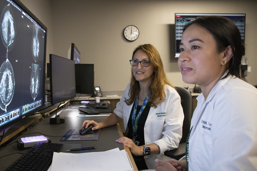
“Our intention is to provide all of our breast patients with the most comfortable environment suited to their individual circumstances,” said Jennie E. Brodsky, MD, assistant professor of radiology and director of breast imaging at the Werths Building. “Patients needing diagnostic imaging may be very anxious and in need of particular care that is respectful of what may be an exceedingly difficult experience. Conversely, those who are coming in for a routine screening mammogram or additional imaging may not have the same needs.”
The Breast Health Center at the Werths Building features four ultrasound rooms, four mammography rooms equipped with the latest Hologic 3Dimensions™ Mammography System, two biopsy rooms, two preoperative rooms and two consultation rooms. Finally, a large reading room is located adjacent to the breast surgery clinic for seamless interdisciplinary care. A testament to thoughtful planning and renowned expertise, the American College of Radiology has already designated the facility as a Breast Imaging Center of Excellence. The center also has capabilities for contrast-enhanced mammography, with biopsy if needed, which can serve as supplemental screening for patients with dense breasts and for patients with an elevated lifetime risk of breast cancer who are unable to undergo a breast MRI.
Thanks to the way the space is configured, patients can easily move between the breast surgery clinic and Breast Health Center without the need to walk through public areas or backroom operations. “To the best of our ability, we accommodate patients so that they are scheduled to see the breast surgeon, to see us in imaging and to schedule any procedures needed — all at the same time,” said Brodsky. “Should it be determined they need a breast biopsy, we try whenever possible to offer that procedure the same day they are here.”
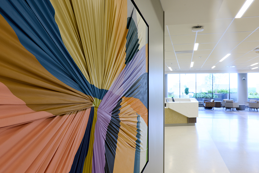
Beyond a cutting-edge spec sheet and renowned specialists across various types of cancer, Mellnick believes that the center’s ambiance — with floor-to-ceiling windows, calming music and bright, open spaces — creates a reassuring environment for patients.
“As patients sit in the light-filled, spacious radiology waiting area and absorb the view of the surrounding neighborhood,” he said, “we hope the feeling of being in a clinical setting is dispelled and they feel a bit more comfortable.”
Published in Focal Spot Fall/Winter 2025 Issue