Song Lab

Projects

Virtual Histology for Assessing MS Pathologies

Goal

Since MRI does not distinguish inter- from intra-axonal water signals, it reflects a weighted-average between inter- and intra-axonal signals. In the presence of inflammation-associated edema or minor axonal loss in people with multiple sclerosis (pwMS), the longer diffusion time for human scanners coupled with the increased inter-axonal space will lead to increased DBSI-lǁ masking the detectability of axonal injury. Thus, through separating inter- and intra-axonal water compartment signals, the sensitivity and specificity to axonal injury of DBSI-derived intra-axonal l|| (DBSI-IA-l||) may be improved. This new model will still preserve the isotropic diffusion specificity to inflammation and tissue loss.

Multiple sclerosis (MS) is an inflammatory demyelinating disease with, ultimately, irreversible axonal injury leading to permanent neurological disabilities. Preventing disease progression or treating progressive MS remains a major unmet clinical need. We propose to: (1) perform DBSI and DBSI-IA analyses on autopsy specimens from pwMS followed by conventional histology and immunohistochemical staining; (2) perform DBSI and DBSI-IA modeling on perfused frog sciatic nerve with and without contrast agent to separate inter-/intra-axonal space water signal; (3) develop a Diffusion Histology Imaging (DHI) approach combining DBSI/DBSI-IA metrics and machine/deep learning algorithms to recapitulate histology specificity to MS pathology; and (4) translate DBSI-IA model to analyze existing DWI data from the cohort of pwMS previously imaged in an expired program project.

Imaging Optic Nerve Function and Pathology: From Mouse to Human

Goal

To regenerate neurons and neural connections in the eye and visual system, requires the development of modalities capable of non-invasively imaging neural connections as they are reestablished between the eye and the brain. We have introduced two promising techniques, diffusion basis spectrum imaging (DBSI) and diffusion functional magnetic resonance imaging (diffusion fMRI) for visualizing the pathology and function of the optic nerve in situ. We combine these technologies to deliver a new, diffusion MRI-based method to assess optic nerve anatomy, function and pathology simultaneously in both mice and human subjects. We have this approach by monitoring the progression and/or regression of axonal damage in glaucoma and optic neuritis. 

We will (1) quantify the relationships between diffusion MRI signals, axon number and visual function in an optic nerve crush mouse model, correlating DBSI with histological counts of axon number and diffusion fMRI with visual acuity; (2) perform in vivo experiments and in silico computation (adapting structural information obtained from histology) on the optic nerve crush mouse model to identify a diffusion time optimized for both DBSI and diffusion fMRI and thus distinguish the contribution of restricted isotropic (distant from the axons) and anisotropic (adjacent to the axons) diffusion; and (3) develop and optimize in vivo human optic nerve diffusion MRI protocol and visual stimulation paradigm that can simultaneously visualize optic nerve anatomy, function and pathology in glaucoma and optic neuritis patients.

Assessing Brain Tumor Pathology Using Diffusion Histology Imaging

Goal

Pathological examination following stereotactic biopsy or surgical resection plays a vital role in current clinical decision-making for the management of glioblastomas (GBM) patients, based on the neuropathologist’s recognition of morphological signatures reflecting tumor cells and changes in the microenvironment, including treatment effects, which are characteristics missed by current MRI biomarkers. Hypothesis: Although an image biomarker of GBM needs to be sensitive and specific to tumor-induced structural changes, structural specificity alone is not sufficient to accurately detect and distinguish underlying tumor pathologies.

We have developed a novel Diffusion Histology Imaging (DHI) approach, combining diffusion basis spectrum imaging (DBSI)-derived MR structural metrics with machine learning, to detect, differentiate, and quantify areas of high tumor cellularity, tumor necrosis, and tumor-infiltrated brain in GBM. We hypothesize that DHI will outperform existing clinical MRI sequences in 1) the detection of tumor cell burden in newly diagnosed GBM patients and 2) the discrimination of tumor recurrence vs. pseudoprogression in GBM patients treated with standard-of-care chemoradiation. Our long-term goal is to utilize DHI as a noninvasive imaging biomarker in GBM patients to improve the diagnostic yield of biopsies, better guide the surgical removal and radiation treatment of tumors, and monitor the effectiveness of clinical trial interventions. We will apply DHI as well as clinical MRI to newly diagnosed GBM patients to assess underlying pathologies of high tumor cellularity, tumor necrosis, and tumor-infiltrated white matter, using the gold standard of tissue biopsy through an established image-guided clinical workflow at WUSM. We will also perform longitudinal DHI in patients following standard-of-care chemoradiation until suspected tumor recurrence as detected by clinical MRI. In patients who are clinically indicated to undergo biopsy or surgical resection, image-guided tumor biopsies per our standard clinical protocol will be correlated with preoperative DHI tumor metrics.

Virtual Histopathology for Accurate Diagnosis of Prostate Cancer

Goal

Therapeutic stratification and management of PCa patients relies on time-consuming pathological examination of biopsy specimens based on the grading of Gleason scores that determine whether a clinically significant tumor is present. Thus, the goal of this project is to validate whether the newly developed diffusion histology imaging (DHI), as a diagnostic device, can noninvasively and accurately detect and grade PCa.
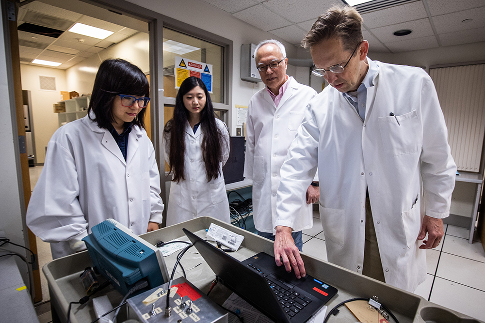
We hypothesize that DHI will accurately detect and assess Gleason scores of PCa eliminating the “blind spots” missed by TRUS-biopsy with the millimeter image resolution covering the entire prostate gland. To prove this hypothesis, we will recruit thirty patients with PSA > 3 ng/mL, scheduled to be biopsied, to undergo DHI and standard-of-care mpMRI measurements of prostate prior to biopsy. Biopsy specimens will be obtained through 1) transperineal template guided biopsy, and 2) mpMRI identified suspicious PCa foci. We will compare DHI-determined Gleason Grade Groups with those determined histologically on the biopsy specimens using confusion matrices and receiver operating characteristic analyses. We will prove the feasibility of DHI-determined Gleason Grade Groups of PCa can accurately reflect histopathological results by transperineal template and TRUS-MRI fusion guided needle biopsy. We will also prove that DHI will fit without interruption of the current clinical diagnostic workflow affording an effective risk stratification and treatment decision for PCa patients.

Our People

The lab, led by Sheng-Kwei (Victor) Song, PhD, includes researchers with expertise in the development of diffusion tensor imaging.