An Lab
Projects
MR Hypoxia Biomarker in Cerebral Small Vessel Disease
In response to the growing societal burden of vascular and other Alzheimer’s related-dementias, NINDS has proposed “Vascular Contributions to Cognitive Impairment and Dementia” (VCID) as a critical area of future research. Cognitive decline in VCID is largely due to progressive cerebral small vessel disease (CSVD). In this grant, we will test a novel neuroimaging biomarker, oxygen extraction fraction (MR-OEF), as a metric of cerebral hypoxia indicating increased risk of CSVD and VCID. By serially imaging adults across a range of vascular risk factors, we will test the ability of MR-OEF to predict neuroimaging progression of CSVD and cognitive decline.
MR Imaging in Sickle Cell Disease
Sickle cell anemia (SCA) is the most common disorder identified on newborn screening, affecting one in 1000 individuals worldwide, with a mean life expectancy of 42 years. Among its complications, stroke and cognitive disability are prevalent in patients with SCA. Our multi-disciplinary team use multi-modal MR approach to study cerebral hemodynamic and metabolic stress, neuroinflammation, and functional and structural impairments.
MarkVCID Consortium
Vascular contributions to cognitive impairment and dementia (VCID) are the second leading cause of dementia, and a major contributor to Alzheimer’s disease (AD), the most common cause of dementia. Non-invasive biomarkers will be critical for the development of prevention and treatment strategies for VCID. A strong partnership between Washington University School of Medicine and the University of Texas Southwestern Medical Center has been developed to serve as a site for MarkVCID Consortium.
3D High-Resolution Cranial Bone Imaging for Pediatric Patients Using MRI
Head CT has been widely used in pediatric patients for the diagnosis of cranial fractures in head trauma and cranial suture patency in craniosynostosis, a congenital disability defined by a prematurely fused cranial suture. Unfortunately, these pediatric patients are particularly vulnerable to ionizing radiation from CT scans. Moreover, sedation is commonly used to reduce motion artifacts in children.
Neuroinflammation in Cerebral Small Vessel Disease using PET/MR Imaging
Cerebral small vessel disease (CSVD) is the leading cause of vascular contributions to cognitive impairment and dementia (VCID). Thus far, the pathophysiology underlying CSVD has not been well-understood. Several lines of evidence suggest neuroinflammation’s potential contribution to the pathogenesis of CSVD and resulting VCID. However, direct evidence of neuroinflammation in patients with CSVD is still lacking.
Image-Guided Noninvasive Stereotactic Cardiac Radiosurgery
Sudden cardiac death (SCD) caused by ventricular tachycardia (VT) results in >150,000 deaths/year in the U.S. Noninvasive Stereotactic Cardiac Radiosurgery (NSCR) was recently developed as a novel noninvasive treatment for treatment-resistant VT, resulting in >95% reduction in VT episodes. In this R01 project, we will implement enhancements to NSCR that will: 1) Reduce the radiation toxicity to the surrounding healthy tissues of the heart and chest to minimize complications from radiation; and 2) Improve the accuracy and efficacy of the radiation dose delivered to the heart lesion that is causing the VT.
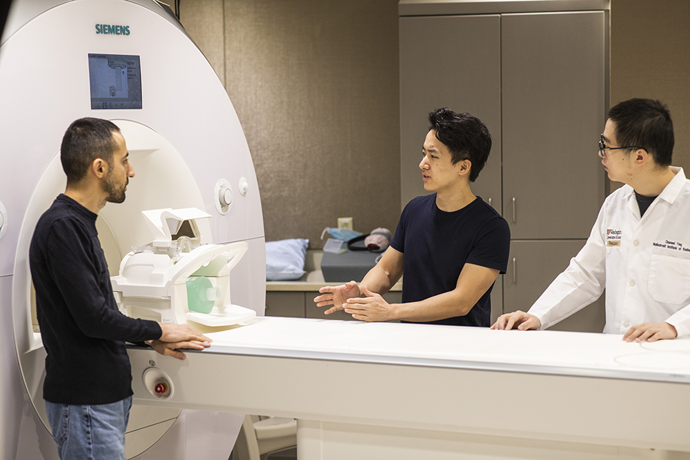
PET/MR Motion Correction and Deep Learning MR Reconstruction
These projects are to develop methods for self-navigated MR and combined PET/MR motion correction. We will also develop novel deep learning-based MR image reconstruction to accelerate MR acquisition.
Our People
The lab, led by Hongyu An, PhD, consists of an interdisciplinary team dedicated in developing MR and PET/MR imaging techniques to impact patient care.