Alumni Spotlight: Cynthia K. Rigsby, MD
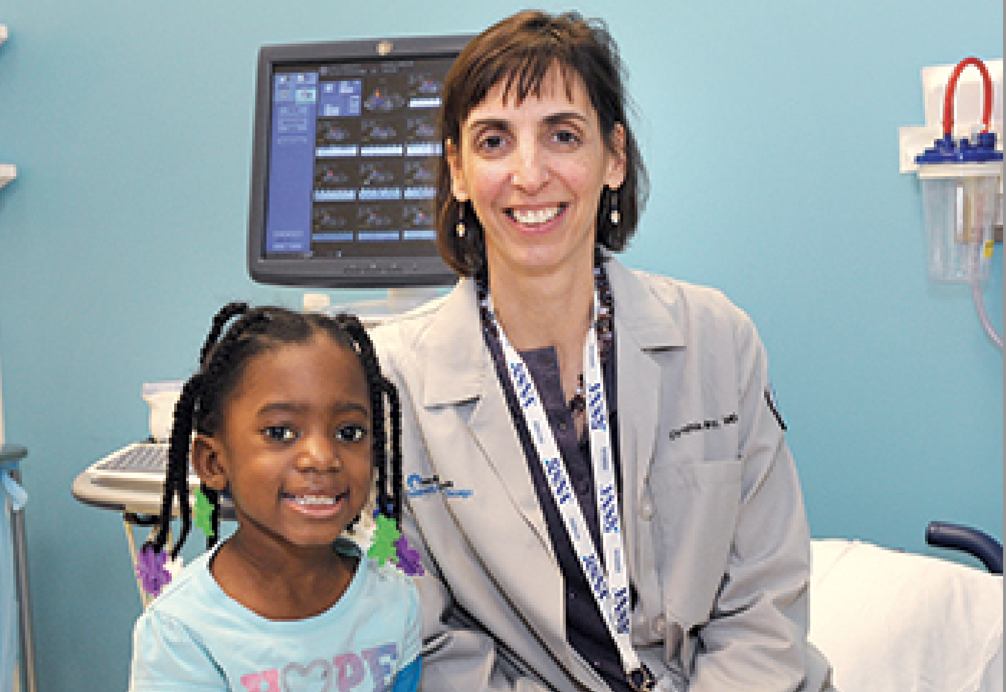
Cynthia K. Rigsby, MD, is a professor of radiology and of pediatrics at Northwestern University’s Feinberg School of Medicine and a pediatric radiologist at the Ann & Robert H. Lurie Children’s Hospital of Chicago (formerly Children’s Memorial Hospital). Rigsby serves as vice chair of the medical school’s Department of Radiology and as division head of body imaging. Inspired by the instruction she received at Mallinckrodt Institute of Radiology during her own residency, Rigsby now uses emerging technology to prepare her own program’s residents for their board exams.
What is your day-to-day life like at Children’s Hospital?
It’s really day-to-day and sometimes hour-to-hour. One-third to one-half of my time is spent doing pediatric cardiac imaging, another 20 to 30 percent is devoted to other pediatric radiology. The rest is administrative time; two areas of focus are overseeing our body imaging protocol and building specialty areas within the body imaging division.
Tell us about the new technology that you’re using with residents.
The American Board of Radiology has changed its board exam from an oral format to a multiple-choice, computer-based exam. That’s very different from communicating with someone in person; we wanted to prepare residents for the new format. I’m using freeware called Socrative, a web-based program that lets you build multiple-choice questions. With a case-based discussion in front of them, the residents log in with their mobile devices and have questions fed to them. They answer and receive immediate feedback, which is nice, and the interactive format helps keep the residents awake! The program tallies all the answers, so as an instructor you can see which questions the residents had trouble with. You can then use that information to fine-tune your own teaching.
Beyond clinical care and administrative duties, what are your research interests?
My clinical and research interests are in pediatric body imaging, especially CT and MRI, with a specialty interest in cardiovascular imaging. Right now, we have a five-year NIH grant to study functional 4D MRI in congenital heart disease. We add live blood flow to a 3D model of the heart to visualize the blood flow within the cardiovascular structure. It really is a tool that provides information that can’t be created any other way.
Last year you were invited back to Mallinckrodt as a visiting professor. How was that experience?
The medical complex has grown enough that I had a hard time getting my bearings! I spent time with Mallinckrodt faculty at St. Louis Children’s Hospital. I also gave a lecture to residents on coronary artery imaging and used the Socrative software to show them some cases. I remember having visiting professors when I was a resident, so I was proud to have been asked to come back.
Who were some of the people at Mallinckrodt who left the greatest impression upon you?
I was attracted to pediatric radiology because of the role models at Mallinckrodt. Marilyn Siegel is the person I wrote my first paper with — a great experience to have as a resident. William McAlister was chief of the section at that time. During case conferences, it was hard to show an unknown case to him that he couldn’t solve. Gary Shackelford was our Teacher of the Year when I was a senior resident, so obviously he influenced many people in our class. He always went the extra step to teach us what we needed to learn, not only at the view box, but with procedures and making sure we had the materials we needed.
Does your time at Mallinckrodt continue to play a role in your professional life today?
Everybody’s roots are in the residency program; it all starts there. From growing up at Mallinckrodt, I have a thirst for knowledge and the desire to teach. We have to pass on the knowledge we have to our trainees. I had a wonderful mentoring program available at Mallinckrodt; I’ve tried to utilize that to figure out how I can mentor others.
You’ve been at Northwestern since 1999, but Children’s Hospital just reopened with a new name and facility two years ago. How has the transition been?
It’s just wonderful. We have bright new private rooms for everyone, with beautiful views of Lake Michigan. We have a state-of-the-art building with all the equipment in imaging we could ever want; I’m like a kid in a candy store. I still can’t believe that when I come to work every day I’m right off Michigan Avenue, and I do a job that I love. Every day I learn something, and I come to work excited about taking care of kids.
You’ve also been credited on several shark research papers. How did that come about?
Years ago I held a joint birthday party for my younger daughter and a kindergarten classmate whose father is a paleontologist specializing in lamniform sharks. He was looking for someone to perform CT scans on a set of rocks that were millions of years old, as well as shark specimens from around the world. So over a couple of weekends we scanned more than 30 shark specimens, including a great white shark head. To this day, he and colleagues are working with the scans of those specimens to sort out all the findings.
What are your interests beyond radiology?
My rare spare time I spend at home with my husband, Michael, and our two daughters: Devyn, 18, and Kristen, 14. I am a gardener; gardening is my therapy. Our home will be part of the garden walk in our community this year. I’m so excited about that; I feel very proud to have my work on display.