Wheelock Neuroimaging Lab
Projects
Network Level Analysis Software
Determining the mechanisms by which the human brain generates cognition, perception and emotion hinges upon quantifying the relationships between coordinated brain activity and behavior. Contemporary connectome research views the brain as a large-scale, complex network composed of nonadjacent, yet connected, brain regions. These connectomes are frequently composed of tens of thousands of connections between brain regions, which poses a challenge for identifying biologically meaningful and reproducible associations with behavioral or clinical outcomes.


Organization of the Developing Connectome
Brain connectivity changes over the course of development to support the acquisition of new skills. Early in development, this includes brain systems supporting primary auditory, visual and motor functions, and later in development, this includes higher-order brain systems supporting complex functions such as language, attention and emotion regulation. Accurately modeling brain development provides valuable insights into healthy and aberrant behaviors. These insights can lead to improvements in early diagnostic and detection tools, a deeper understanding of neuroplasticity and specialization within the brain, and a clearer comprehension of how early experiences impact brain connectivity.

Connectome Degeneration in Alzheimer Disease
In Alzheimer’s disease (AD), a cascade of events — including the accumulation of amyloid-beta (Ab) plaques, neurofibrillary tau tangles, cortical thinning, hypometabolism and disrupted brain connectivity — leads to severe cognitive deficits. Blood-based assays detecting neurofilament light chain (NfL), a structural protein indicating axonal damage, have emerged as potential biomarkers for neurodegeneration and disease progression in AD. Additionally, hub regions that are crucial for efficient brain communication have shown promise as potential neuroimaging markers due to their vulnerability to pathology and degeneration in AD.


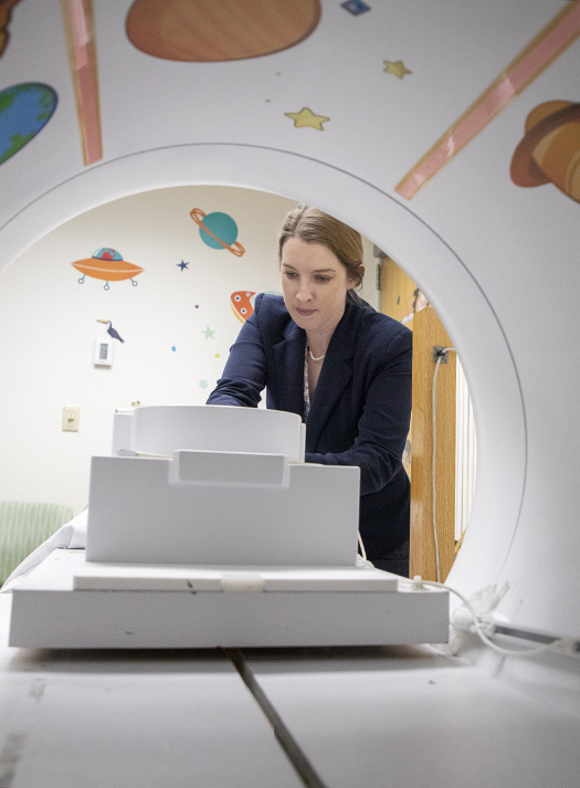
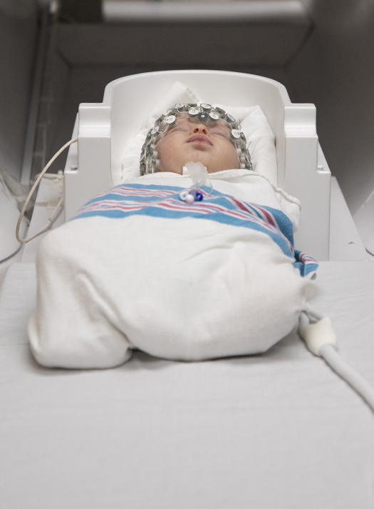
EEG-fMRI
Determining accurate models of the developing brain’s functional architecture during the first two years of life is necessary to make predictions about developmental and clinical outcomes. Crucially, developmental differences in sleep cycles between infants and toddlers and individual variability in time spent in each sleep stage pose a challenge for both understanding early brain development as well as mental health outcome prediction reproducibility and accuracy in extant pediatric, sleeping state fMRI studies. Disentangling the relative contributions of sleep stage and age in early brain network development will be transformative, providing a state-based understanding of early developmental brain networks and a framework for accurate developmental outcome predictions.