Small Animal Magnetic Resonance Facility
Services & Equipment
Scanners
The 4.7-T Agilent MRI scanner is also multinuclear and dual channel, employing horizontal Oxford Instruments (Abingdon, Oxfordshire, UK) magnets of 40- cm clear bore diameter. The 40-cm system utilizes a current generation, actively shielded Resonance Research Inc. gradient/shim coil assembly of 11.5-cm, driven by Oy International Electric Company (IEC; Helsinki, Finland) model A-240 amplifiers.
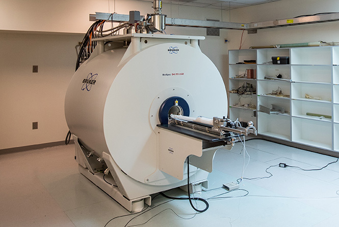
The 9.4-T Bruker MRI scanner, newly installed in the fall of 2020, has a 20-cm clear bore, is multinuclear, and has four receiver channels. The gradient system has a 11.4-cm inner diameter, is driven by IEC amplifiers (500 V / 300 A), and is capable of delivering 66 gauss/cm with a rise time of 70 µs. Notably, this scanner includes a 4-channel array mouse brain CryoProbe which offers substantially improved SNR over traditional RF coils.
The 15.2T Bruker MRI scanner — to be delivered in fall 2025 — has an 11-cm clear bore, is multinuclear and has four receiver channels. The gradient system has a 6-cm inner diameter and is driven by IEC amplifiers (300 V / 200 A).
Space & Additional Equipment
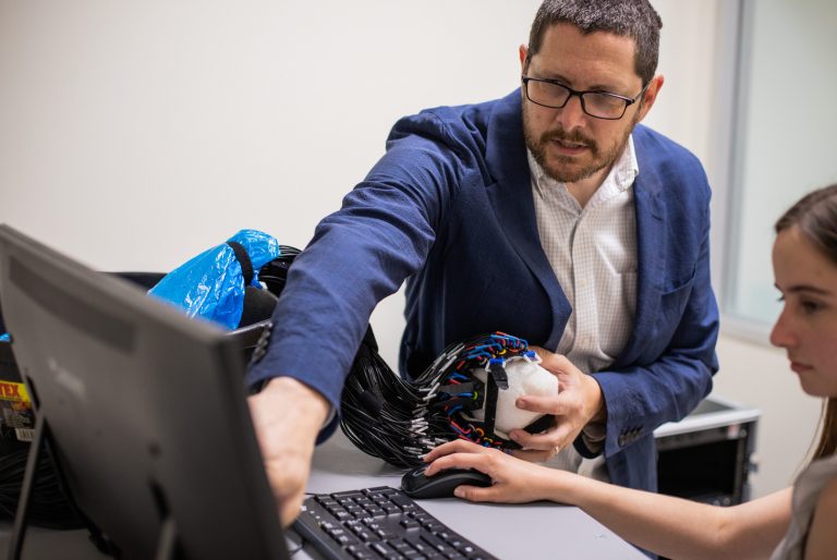
- The Facility maintains a substantial suite of commercial (manufactured by Stark Contrast, Agilent/Varian, Bruker, and Doty Scientific) and laboratory-constructed RF coils. These include a wide array of both quadrature volume coils and surface coils. In particular, the Facility has developed substantial experience and expertise in performing MR experiments using actively-decoupled surface (receive) / volume (transmit) coil pairs. This coil arrangement takes advantage of the high sensitivity of the surface coil and the excellent RF homogeneity of the volume coil.
- The 9.4T Bruker MRI system supports actively decoupled receiver coils with up to four channels. Currently available multi-channel array coils on that system include a 4-channel rat brain surface coil, a 4-channel mouse brain surface coil and a 4-channel mouse brain CryoProbe, which offers a 2-3x increase in SNR over our more traditional ‘room temperature’ 4-channel array coil.
- The 15.2T Bruker MRI system supports actively decoupled receiver coils with up to four channels. Available multi-channel array coils on that system will include 4-channel mouse head and mouse body array surface coils.
Across the hall from the surgical suite are two dedicated, specially-designed, 150-sq-ft holding rooms for animals. These small-animal quarters are managed by the Division of Comparative Medicine. The University also maintains state-of-the-art barrier and non-barrier animal facilities immediately across the street. A modern histology facility, outfitted with equipment for intracardiac animal perfusion, embedding brain tissue in paraffin or plastic, tissue sectioning, tissue staining, and histologic analysis (either by electron microscope, light microscope or computer-aided image analysis) is also located nearby.
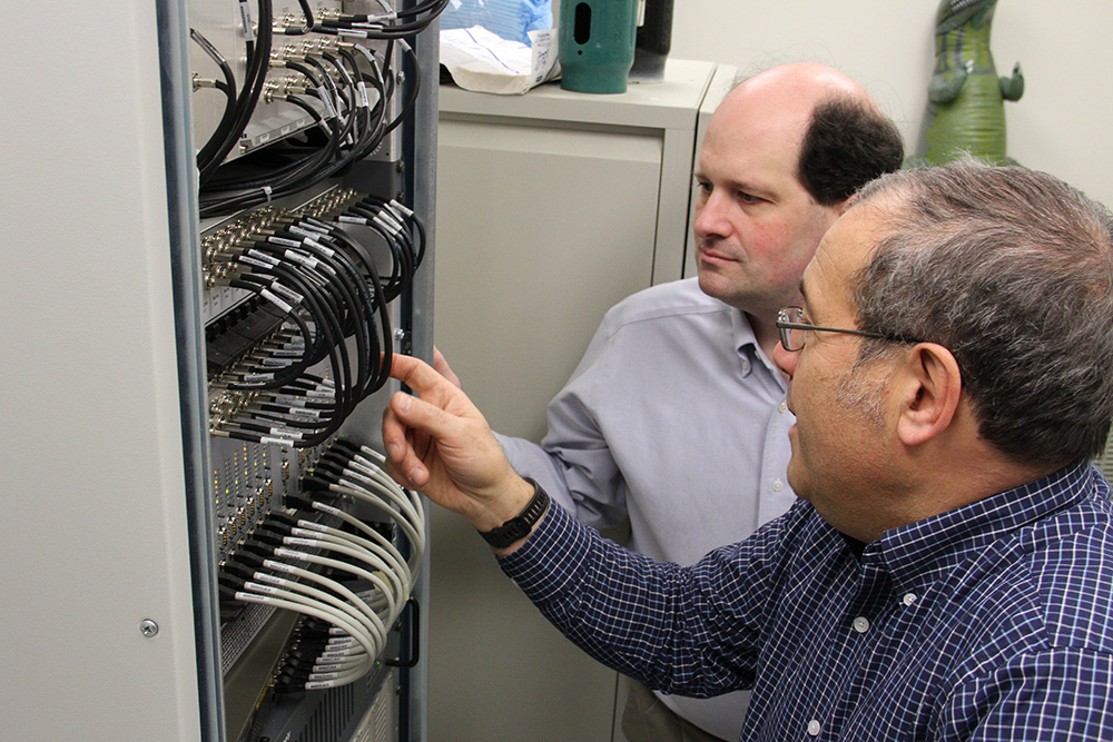
Wet-chemical and electronic labs are located on either side of the surgical suite. The 200-sq-ft wet lab is equipped with a refrigerator/freezer for storage of compounds, an analytical balance, and a centrifuge. The 200 sq ft electronic lab, which includes RF oscilloscopes and a network analyzer, is well equipped for coil design and spectrometer modification.
Computational Software
- MATLAB
- Mathematica
- In-house developed Bayesian analysis toolbox
- Sun (Solaris) and Intel (Linux) compilers for parallel programming in OPENMP
- Analyze
- ImageJ
- ITK-SNAP
Hyperpolarizers
A commercial GE SPINlab DNP hyperpolarizer instrument is installed in the East Building in close proximity to the Siemens 3T and MR Solutions MRI systems.
- The QC Accessory Module is equipped for rapid assays on samples for human injections.
- A sample drop-chute located immediately adjacent to the polarizer provides access to preclinical MRI scanners located one floor below.
- Operates at a 5T magnetic field, 0.8 K temperature, and 140 GHz microwave irradiation to generate hyperpolarized 13C/15N MRI contrast media with polarization levels approaching 60% or ~5 orders of magnitude higher than possible using standard magnets.
- Sterile pharmacy kits are filled with 13C-enriched pyruvate in a biosafety cabinet, in collaboration with the Biologic Therapy Core Facility (BTCF).
- After loading into the SPINlab and a polarization buildup time of ~3 hours, each sample is rapidly dissolved by a bolus of superheated water at 130° C / 250 psi.
- The SPINLab can accommodate up to four samples simultaneously, with dissolutions spaced as little as 5 minutes apart.
Contact: Cornelius von Morze, PhD
The NVision POLARIS system is expected to be delivered in fall 2025.
- Enables hyperpolarization of [1-13C]pyruvate with high throughput by automating the method of PHIP-SAH (parahydrogen induced polarization with side arm hydrogenation), achieving levels of polarization and purity that rival DNP
- Works at room temperature without need for continuous chilled water supply and other infrastructure adjustments
- Can dispense doses of HP [1-13C]pyruvate every few minutes, enabling much higher throughput than DNP
- Additional probe capabilities currently under development
Contact: Caroline Guglielmetti, PhD
Contact Us
For more information or to start a new project, email James Quirk, PhD.