Hershey Lab

Projects

The goal of the Wolfram Clinic is to better understand the development and progression of the neurological aspects of Wolfram syndrome. During annual research visits patients are seen by specialists in multiple disciplines (ophthalmology, neurology, psychiatry, audiology) and undergo magnetic resonance imaging (MRI) of the brain. This data helps us understand the neurological issues in Wolfram syndrome, and will directly inform the design and metrics used in future clinical trials.
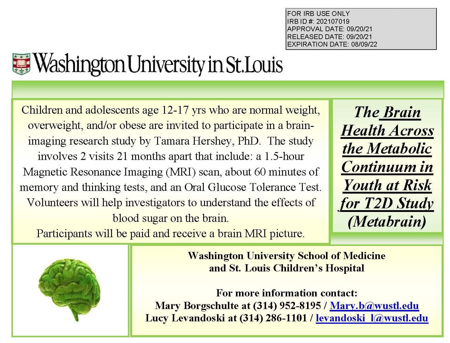
Wolfram syndrome is a rare genetic (autosomal recessive) disease originally described as the combination of insulin dependent diabetes mellitus, optic nerve atrophy, diabetes insipidus and deafness. Neurodegeneration and neurological features were thought to appear at later stages of the disease, ultimately leading to death in middle adulthood (Barrett et al., 1995). There are no interventions to slow or stop this devastating deterioration.

Funding: Hershey, PI
(R01 HD070855) 2012-2023
HRPO #201301004

See a list of publications resulting from the Wolfram Clinic.

Learn more about the Wolfram Clinic, including information about:

  • Wolfram Family Conference
  • Biannual Clinic Newsletter
  • Clinical Care and Consultation
  • How to Participate in a Study
Pons volume in Wolfram syndrome (colored dots) is highly discrepant from controls (gray diamonds)

Type 2 diabetes mellitus (T2D) is a significant public health problem affecting ~30 million Americans. Obesity, insulin resistance, insulin deficiency (β cell dysfunction) and dysglycemia all precede the diagnosis of T2D and are known to promote inflammation and ultimately lead to microvascular complications. More recently, research has identified brain-related complications in adult-onset T2D, including reduced regional brain structure and function, impaired cognition, and increased lifetime risk for Alzheimer’s disease. Alarmingly, an increasing number of children and adolescents are being diagnosed with T2D, likely due to the growing prevalence and earlier onset of obesity. Youth-onset T2D appears to have a more aggressive course than adult-onset T2D, with earlier onset and more rapid progression of microvascular complications. In addition, studies of youth with obesity and youth-onset T2D have reported robust differences in regional brain structure and cognition, suggesting that brain effects may follow the same aggressive course as the more typical vascular complications. Unfortunately, little is known about the factors associated with poor brain structure and function in youth with T2D.

To address this critical gap in knowledge, we propose to study youth across the spectrum of body mass index (BMI) and metabolic dysfunction. This approach will allow us to disentangle the relationship of key features of T2D risk (e.g. obesity) with intermediary physiologic changes that pose a risk for the brain (e.g. insulin resistance, inflammation, β-cell dysfunction and dysglycemia) that may lead to reduced brain structure and function in T2D. We will determine which of these factors are most associated with differences in brain structure and function among groups, over time, and how these effects differ from normal neurodevelopment. Given that the disease occurs at a time when brains are undergoing dramatic developmental processes, the aggressive nature of youth-onset T2D progression and complications in other organ systems, these results may provide guidance and justification for longer follow-up, interventional or mechanistic studies and have important clinical implications. 

Hershey, PI
(R01 DK126826) 2021-2026
HRPO #202107019

The Brain Health Across the Metabolic Continuum in Youth at Risk for T2D Study (MetaBrain):

Children and adolescents age 12-17 yrs who are normal weight, overweight and/or obese are invited to participate in a brain imaging research study by Tamara Hershey, PhD, The study involves 2 visits 21 months apart that include: a 1.5-hour magnetic resonance imaging (MRI) scan, about 60 minutes of memory and thinking tests, and an Oral Glucose Tolerance Test. Volunteers will help investigators to understand the effects of blood sugar on the brain. 

Participants will be paid and receive a brain MRI picture. 

Washington University School of Medicine and St. Louis Children's Hospital

For more information, contact:
Mary Borgsculte at 314-952-8195 / mary.b@wustl.edu
Lucy Levandoski at 314-286-1101 / levandoski_l@wustl.edu

The Hershey Lab and collaborators are studying how the brain is a target organ in diabetes. In particular, we are interested in how brain development and exposure to hyperglycemia and hypoglycemia interact to shape developmental trajectories of the brain and its functions. This work has led to an understanding of the regional brain vulnerability in diabetes and has been funded for the past 10 years by the NIH (NIDDK).

Enrollment for study has ended; analyses are ongoing.

The purpose of the NewT research study is to determine how symptoms at the time of Type 1 Diabetes Mellitus (T1DM) diagnosis, such as hyperglycemia and diabetic ketoacidosis (DKA) interact with age of onset to shape the development of the brain.

Funding: Hershey, PI
(R01 DK064832) 2016-2021
HRPO #201601135

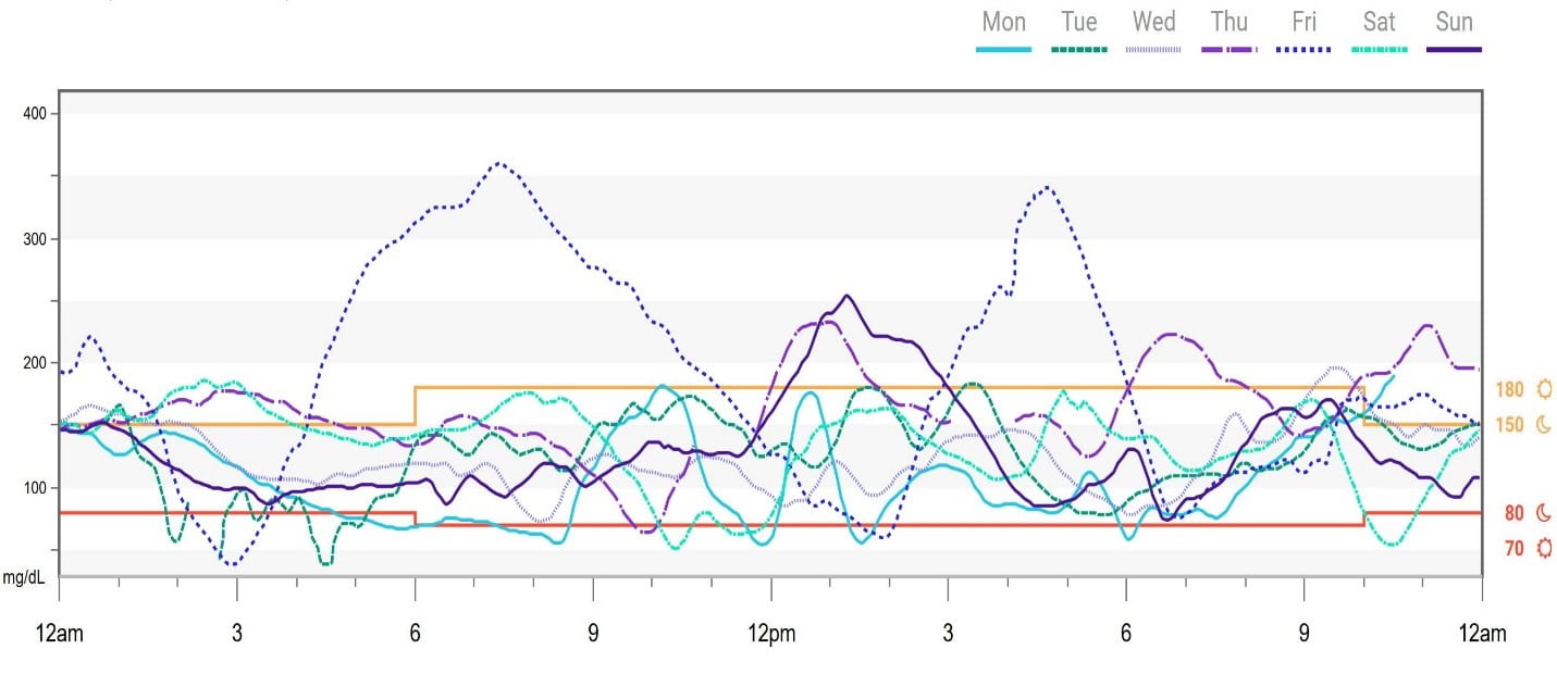
Management of blood glucose with type 1 diabetes (T1D) is challenging for youth and often results in frequent swings between normal, high, and low glucose (Awoniyi, 2013) like that shown in Figure 1. This glucose variability remains poorly understood, but research has shown that it is a potential risk factor for diabetic complications (Ceriello, 2019). One of the least understood complications of T1D is cognitive impairment. Several studies have shown impaired cognitive function in youth with T1D compared to their non-diabetic peers, and that poorer glycemic control including severe glycemic events (e.g., severe hypoglycemia) and chronic hyperglycemia are associated with cognitive impairments (Cato 2016). These studies assessed the important relationship between long-term glucose variability in T1D and cognition; however, little is still known about how cognitive function is impacted daily in real-life settings, particularly with dynamic cognitive skills that fluctuate throughout the day (e.g., working memory) (Gamaldo, 2016).

The goal of the PARC study is to evaluate real-time, real-life dynamic cognitive function variability in youth with T1D using a unique combination of novel, continuous health methods (i.e., continuous glucose monitoring), cognitive data collection methods (i.e., smartphone application), and machine learning models. Understanding how these dynamic cognitive skills are affected in daily life could be an important first step in establishing crucial improvements in academic accommodations and treatment recommendations for youth with T1D.

An example of typical daily glucose fluctuations from continuous glucose monitoring in a patient with T1D in good control (HbA1c=5.3%)

Obesity is a major risk factor for diabetes and late-life cognitive impairment and dementia, including Alzheimer disease. As longevity rises and the rate of obesity increases globally in children and adults, it is important to understand mechanisms by which obesity confers risk for late-life disease. One such mechanism may be obesity-related brain inflammation, or neuroinflammation. 

Animal models of obesity show that overfeeding causes neuroinflammation throughout the brain accompanied by deficits in hippocampal-dependent memory tasks. In humans, noninvasive yet in vivo magnetic resonance imaging (MRI) allow us to investigate putative neuroinflammation in humans with obesity relative to those with normal weight. We have observed greater putative cellularity and reduced putative axonal and dendritic densities, as reflected by the diffusion MRI method diffusion basis spectrum imaging (DBSI), in white matter tracts, hippocampus, hypothalamus and nucleus accumbens in children and adults with obesity relative to their normal-weight peers. These findings indicate that obesity may be associated with neuroinflammation relative to normal weight. In addition, DBSI metrics relate to body mass index, self-reported feeding (e.g. emotional eating) and cognitive function. Current and proposed studies aim to establish reliability of DBSI metrics in humans with normal weight and obesity over time and determine whether putative neuroinflammation in obesity and co-occurring insulin resistance and cognitive dysfunction can be mitigated by weight loss. Results of these studies will indicate whether obesity-associated neuroinflammation is a target for prevention or treatment of obesity-related conditions including cognitive dysfunction and late-life cognitive impairment and dementia.

Sarah A. Eisenstein, PI
2022-2023: Mallinckrodt Institute of Radiology Pilot Funds and McDonnell Neuroimaging Labs (NIL) Innovation Fund
IRB# 202112165

2018-2022: Diabetes Research Center Pilot & Feasibility Award
IRB# 20105053

Human obesity is a major public health problem that is driven by a complex combination of behavioral, genetic, environmental, biological and neurobiological factors. Neuroimaging studies in humans find altered dopamine (DA) function and reward-related behavior associated with obesity but conflicting findings limit our understanding of these complex relationships.

The Hershey Lab has investigated striatal D2 dopamine receptors in non-diabetic obesity and found that D2R binding was related to obesogenic eating, reward-related traits and genetic markers of DA signaling across both obese and normal weight subjects. Furthermore, we found that lower glucose-induced pancreatic insulin release in non-diabetic subjects related to altered reward-related traits. Together, these data suggest that dopamine, insulin and reward may be linked in ways that help us understand neurobiological risk for and response to obesity.

Proposed model of interactive factors linking genetics, D2R, insulin resistance and behavior in human obesity.

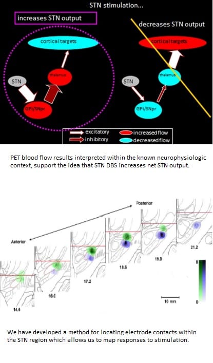
Deep-brain electrical stimulation (DBS) can provide substantial motor benefit and impact non-motor domains of cognition and mood in Parkinson’s disease (PD) when implanted in the subthalamic nucleus (STN). However, the neural mechanisms underlying these effects are not fully understood. We are collaborating with the Culver Lab to use high density diffuse optical tomography (HD-DOT) to understand how STN DBS alters functional cortical networks in the brain.

Funding: Hershey/Culver, Co-PIs
(R01 NS109487) 2019-2024
HRPO #201908154

An example of typical daily glucose fluctuations from continuous glucose monitoring in a patient with T1D in good control (HbA1c=5.3%)

More in Hershey Lab

Latest in the NIL-RC