Gordon Precision Neuroimaging Lab
Projects
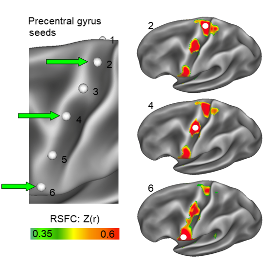
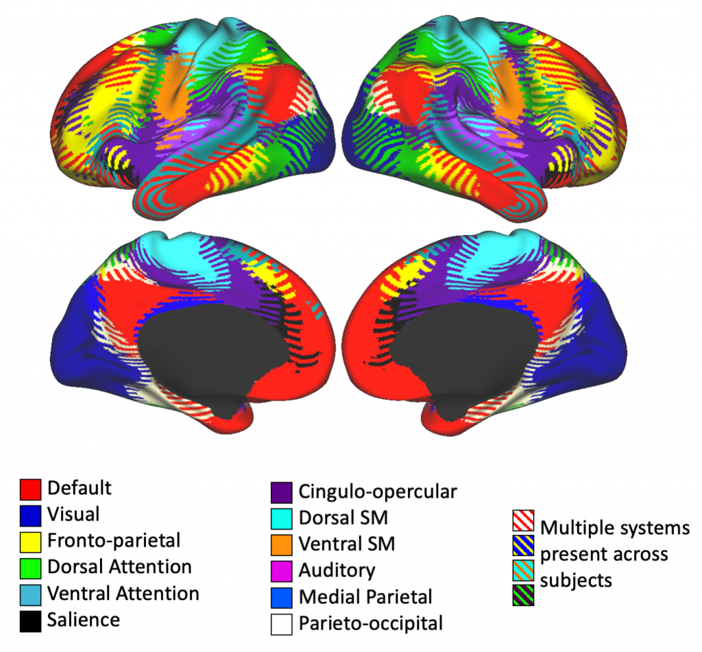
Understanding and Mapping Novel Aspects of Human Brain Organization
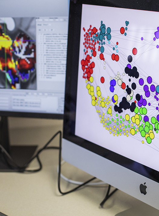
The human brain is organized into a series of large-scale brain networks that enable sensation, action, and complex cognition. We aim to map these brain networks in the finest possible detail, including their relationship with brain structure and their representation in cortex and subcortical structures. We are able to identify new features of brain network organization that have never before been characterized.
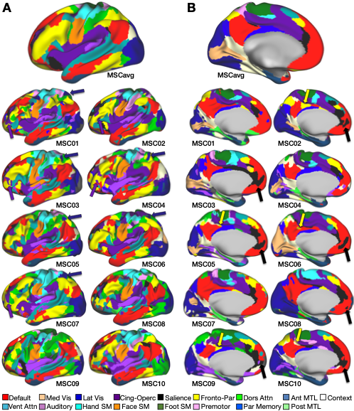
Individual Variability in Brain Organization
Individuals exhibit substantial inter-individual variability in the size, location, and connectivity of their brain networks. This project aims to characterize those differences, and ultimately to link variability in brain networks to individual differences in personality and mental abilities.
Methods Development for Precision Neuroimaging
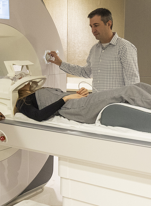
We are actively developing novel approaches to precisely map functional brain networks in individual humans. These efforts include characterizing, modeling, and predicting the within-individual reliability of brain network measures; improving mapping of brain networks into deep structures including the diencephalon and brainstem; and mapping brain networks at high resolution using 7-tesla imaging.
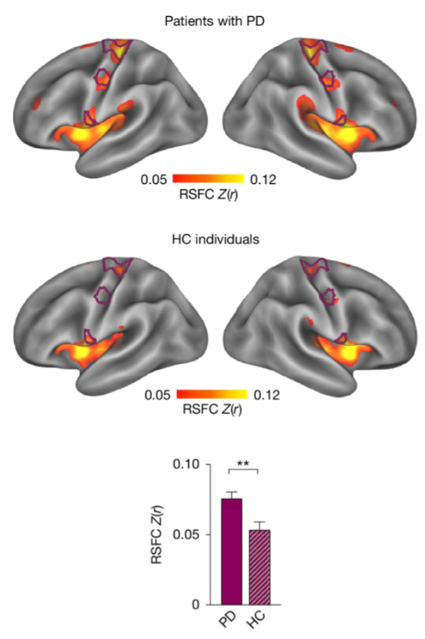
Precision Neuroimaging of Parkinson’s Disease
Parkinson’s Disease is classically considered a “movement” disorder, yet Parkinson’s symptoms also include deficits in initiating actions, continuing actions once begun (e.g. “gait freezing”), and motivation. We are testing whether specific brain circuits we have identified in healthy controls that link motivation to action to movement may explain the range of Parkinson’s symptoms. We are further evaluating whether these circuits may serve as ideal targets for neuromodulation therapies.
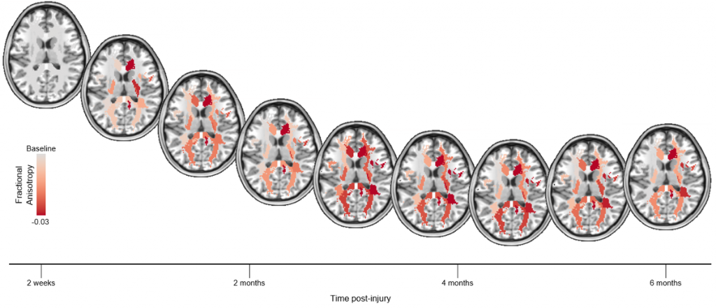
Brain Network Changes After Traumatic Brain Injury
Traumatic brain injuries damage the white matter fibers linking networked brain regions, disrupting brain processing and affecting cognition, emotion, and sensation. In many patients, recovery from a TBI can take months or even years. We are using neuroimaging to longitudinally track patients who have suffered from a TBI to better understand how these recovery processes play out in the brain.
Neuroimaging of Dystonia
Dystonia is caused by abnormal brain function in circuits linking the cerebral cortex to the striatum and cerebellum. We are precisely mapping those circuits in individual patients and characterizing how a common treatment for dystonia alters their function and their connectivity.