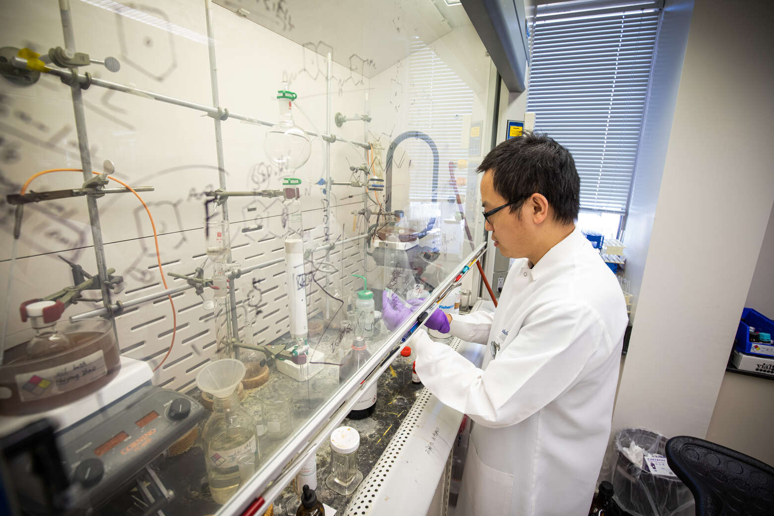
Do You Know the Differences Between a CT, MRI and PET Scan?
You’ve heard about them. You may even have had one or more of them. But do you really know the difference between these imaging tests? Most people don’t, nor do they understand that a radiologist is a “real” doctor working in the background to make a diagnosis.
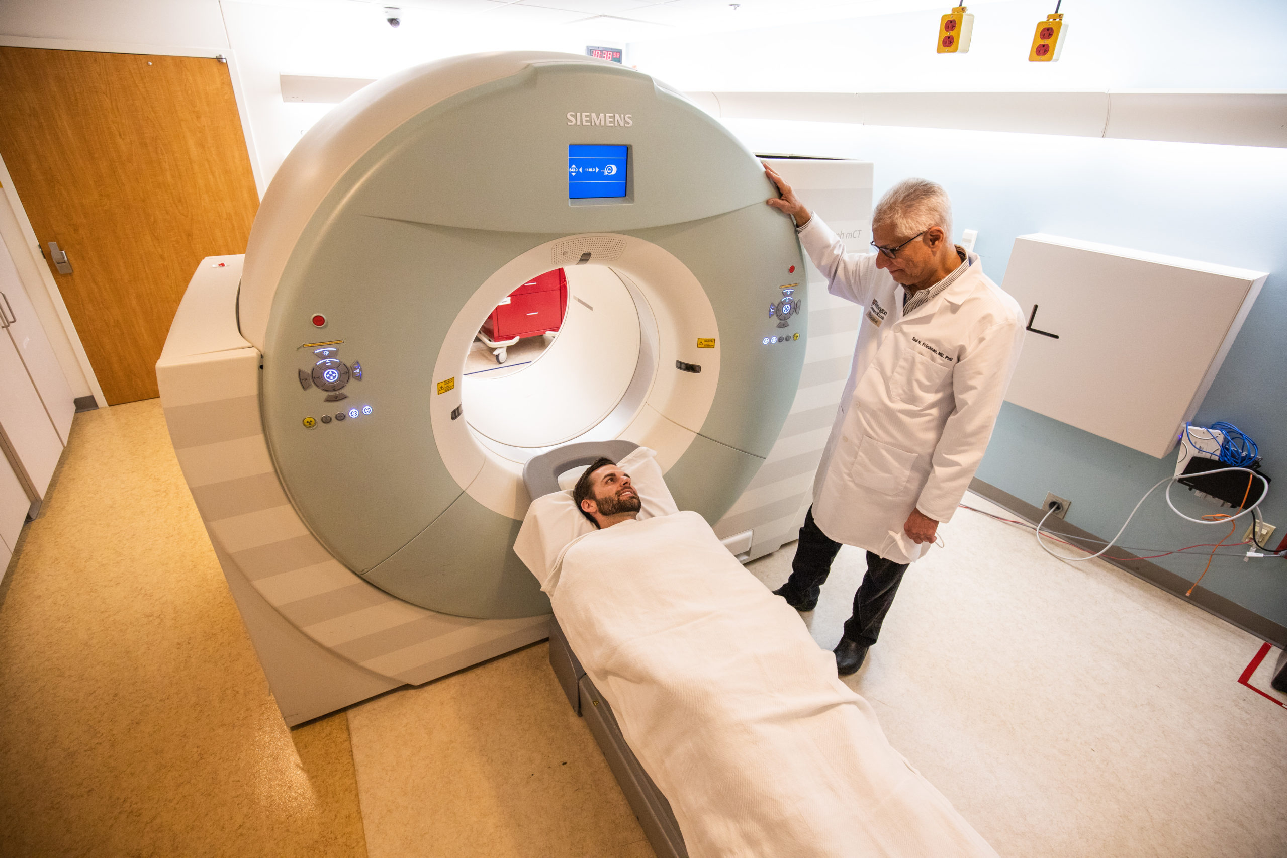
CT (computed tomography)
This imaging exam uses a thin X-ray beam to produce detailed images inside your body. It takes multiple cross-sectional images to show the “shape” of internal organs and other structures, such as bones and blood vessels. The images are then re-assembled, like a loaf of bread, and displayed on a computer monitor for a radiologist to “read” or interpret. The image can be “spun” allowing the radiologist to view the organ or structure in question from various angles.
A CT shows more detail than an X-ray and is used, among other things, to diagnose cancer, heart disease, injuries from trauma, and musculoskeletal disorders. It’s one of the most common imaging tests used today. Just like X-rays, a CT uses radiation to acquire its images. Because they are quick, CT scans are often used in emergency departments.
MRI (magnetic resonance imaging)
Unlike a CT scan which uses radiation, an MRI uses a powerful magnet and radio waves to create detailed images of organs and other structures inside your body. Consequently, patients undergoing this test must remove metal items and tell the technician if they have any internal devices (such as a pacemaker, stent or artificial joint) that contain metal. This test is quite noisy.
An MRI is used to diagnose a variety of conditions from torn ligaments to tumors. It provides different information from a CT. Some MRI exams require a contrast agent (a dye) that’s injected through an intravenous (IV) line to better view problem areas. An MRI takes longer to perform than a CT and is more expensive, but it doesn’t expose you to radiation.
PET (positron emission tomography)
A PET scan is used to see metabolism (chemical activity) within your body. It can detect abnormal changes before structural changes occur. Said another way, a PET scan can detect cancer before a tumor forms or can be “seen” on a CT or MRI scan.
A PET scan involves injecting a very small dose of a radioactive substance through an intravenous (IV) line. As the radioactive substance (also called a radiotracer) courses through your body, the PET scanner detects and records its activity. A PET scan is used to diagnose cancer, heart disease, and some brain disorders. It supplies information that’s different from an MRI or CT scan. While a CT and MRI scan looks at the “form” of structures inside your body, a PET scan looks at their function and shows unusual cellular activity.