

From Bitter Pill to Gummy Vitamin?
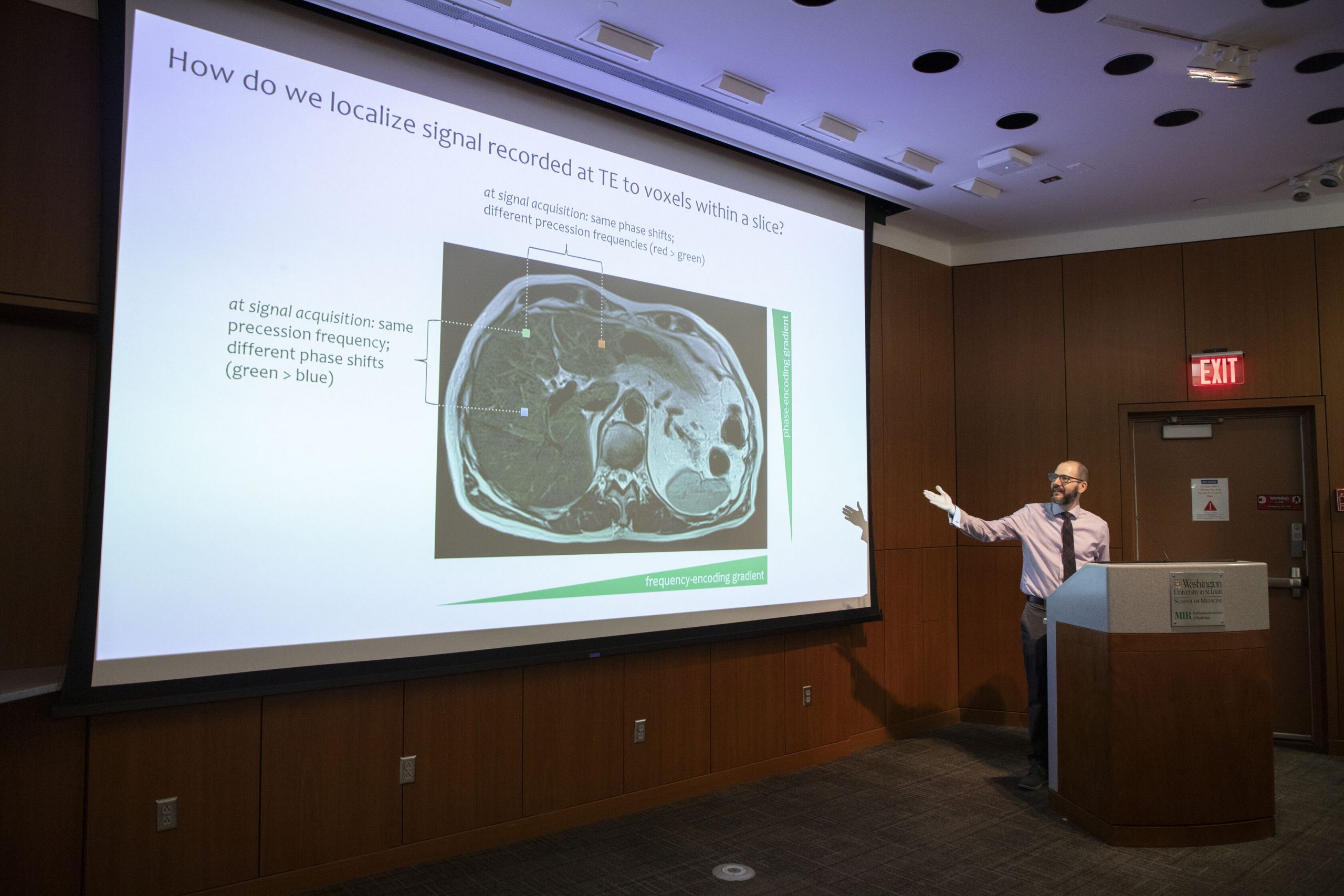
A crucial but often dreaded subject in radiology education is medical physics. Often considered a “bitter pill” that residents must swallow to complete their studies, physics curricula are notoriously dry and can sometimes obfuscate the link between abstract theory and daily practice. But where many saw a dense lesson plan, Tyler J. Fraum, MD, associate professor of radiology, saw an opportunity to turn this bitter pill into a cognitively nourishing yet enjoyable “gummy vitamin.”
MIR’s previous physics curriculum covered nuclear and non-nuclear medicine topics over two years; however, the order and complexity of the lectures didn’t align with the academic calendar, leaving new residents sitting through advanced lectures without foundational knowledge. When this perennial complaint resurfaced, Fraum and other physics-focused individuals in the department banded together to revitalize the curriculum and improve physics education at MIR.
Reshaping 64 hours of content was no small feat. Fraum knew he needed support to complete the project, which he found in the Loeb Teaching Fellowship through the WashU Medicine Academy of Educators. The two-year program provides protected time to develop and implement new medical education ideas at the university. “I didn’t want the momentum that we had initially to be lost,” he said.
The new curriculum prioritizes teaching the foundations of radiologic physics in a logical sequence before diving into more advanced subjects, organizes content around the individual modalities and emphasizes clinical application. Other changes include more active learning strategies, such as interactive polls that ask questions and enable real-time responses and discussion. For the nuclear medicine portion of the curriculum, Fraum delivers six board review-style lectures that revisit earlier lessons for those about to take their core exams. Another addition, an annual introductory physics “boot camp,” was inspired by resident listening sessions. Held in July, the boot camp ensures that trainees understand the basic principles of radiologic physics, no matter when they enter the program.
Ultimately, the goal is to help residents connect how physics impacts their day-to-day work. “I try to make the physics as clinically relevant as possible,” he said, “so that residents appreciate why they need to know what we’re teaching them and how these concepts can be implemented in their actual practice as radiologists.”
“We want to measure effects of the curriculum with the goal of publishing the MIR experience.”
Now nearly halfway through the Loeb fellowship, Fraum continues to hone the curriculum and plans to add more active learning strategies. He has also been collecting quantitative data to determine the curriculum’s efficacy, not only to enhance the next iteration but to help grow communal knowledge of radiologic physics education. “It’s not just about local implementation,” said Fraum. “We want to measure effects of the curriculum with the goal of publishing the MIR experience to help other programs learn from our challenges and successes.”
Making Sense of Medical AI
Nearly every facet of modern radiology — from image generation to workflow management and reporting — has been touched by artificial intelligence (AI). As more tools are introduced and integrated into medical institutions, educators like Shinjini Kundu, MD, PhD, assistant professor of radiology, are now tasked to prepare trainees to practice radiology in the era of AI.
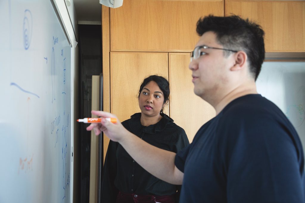
Kundu, also a principal investigator in the Computational Imaging Research Center, has developed a new resident curriculum dedicated to AI and medical imaging. So far, the curriculum includes two dedicated noon conference lectures a year that use real-time quizzes for a more active learning experience. The core content covers basic AI definitions, existing tools and their uses across fields; however, Kundu said she wanted to go beyond the basics and ground the lessons in what would be useful in their practice. She teaches how machine learning models are trained, tested and validated; how to discern between well-crafted and inferior models; and how cognitive biases and personal trust in AI tools affect their efficacy. “It’s not simply about the tool. It’s about the complex ways a human interacts with that tool,” she said.
Not all AI tools are created equal, and for radiologists, it’s important to understand how a tool’s limitations and nuances impact patient care. “Many of these tools are designed for a narrow context,” Kundu said. “While these models are trained on specific data, they cannot always account for the nuanced needs of the patient in front of you. Advocating for a patient’s best interest is something only a human physician can provide.”
“Advocating for a patient’s best interest is something only a human physician can provide.”
She doesn’t expect residents to become AI experts or develop their own models, but some residents have gone above and beyond. Ramanan Sivakumar, MD, a diagnostic radiology resident, reached out to Kundu in his first year about pursuing a research project on AI. They created a study to examine the current landscape of AI tools that receive clearance from the Food and Drug Administration. “I believed it would be beneficial for him to understand how the FDA carries out its process because he will probably encounter FDA-cleared tools in clinical practice,” Kundu said.
Sivakumar pulled data from nearly 1,000 radiology-specific AI and machine learning vendors that submitted applications for FDA clearance and compared whether they underwent clinical testing or not. Since completing the study, Sivakumar has published his findings in the Journal of American Medical Association Network and presented at the Association of Academic Radiology.
Training for the Unknown
Complications in medicine are inevitable, so how do educators prepare their trainees for the unknown? Arindam R. Chatterjee, MD, associate professor and director of the neuroendovascular intervention fellowship program, has developed a course to give neurointerventional fellows the opportunity to gain deliberate practice in managing procedural complications.
Fellows Leaping Into Practice (FLIP) is a three-day radiology oral board-style course focused on navigating complications and receiving immediate feedback. While fellows encounter numerous successful cases in their training and have access to educational content, Chatterjee said it can be challenging to capture the high-stress demands of rare real-life case complications.
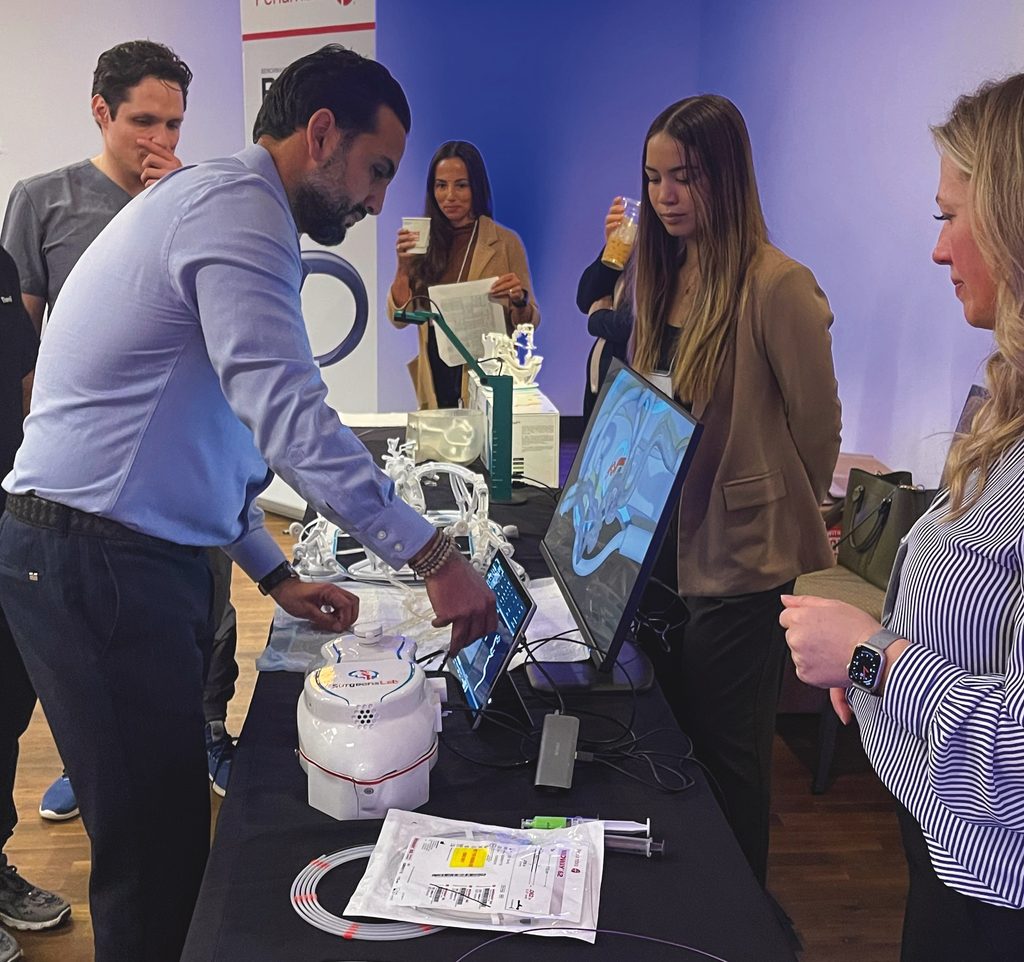
FLIP launched in 2022 in partnership with Penumbra, a medical device company. Core faculty are from MIR, the Departments of Neurosurgery and Neurology, and additional nationally recognized faculty from around the country. FLIP sessions invite fellows to share cases and complications they’ve experienced in their training with a panel of faculty members in various disciplines. The panel discusses how the cases went and different approaches to take in the future — everything from preventing the complication to managing it in the moment.
Another session involves simulation cases in a style inspired by the radiology oral boards. Chatterjee described this as an interactive “choose your own adventure” exercise where students must determine how to proceed, and mentors provide immediate, specific and actionable feedback modeled after Anders Ericsson’s work on deliberate practice. “When they’re sitting one-on-one, the students are the ones doing all the talking, and the mentor is there to guide them through the case. If they give the wrong answer or an answer that would lead to a complication, then the mentor may say, ‘The patient died. Do you want to start this case over?’”
FLIP was first held in St. Louis, but Chatterjee has taken the program on the road to Houston, Washington, D.C., and New York City. Now in its fifth year, the course is not only unique, but popular. “It’s quickly become a high-value educational resource with about 60 participants per year, and it typically has a waitlist,” Chatterjee said. “We’ve had so much enthusiasm that graduates are asking to return as faculty.”
About 800–1,000 neurointerventionalists practice in the U.S. — a small but impactful subspecialty. Many medical facilities have only one or two neurointerventional radiologists on staff, and even practitioners at larger academic institutions rely on their fellow practitioners for guidance. Chatterjee said an added benefit to the course is building a network of practitioners who contribute to the field’s knowledge pool and can serve as trusted resources. “All of us encounter complications in this field that
continues to evolve at a rapid pace,” he said. “While complications are rare, new devices can lead to situations we haven’t encountered before. By openly sharing our experiences and discussing complications as a field, we can collectively advance care and improve outcomes for patients everywhere.”
Published in Focal Spot Spring/Summer 2026 Issue